

Method for quickly, directly and directionally inducing differentiation from mouse embryonic stem cells to neuroepithelial cells
A technology of neuroepithelial cells and embryonic stem cells, applied in animal cells, nervous system cells, vertebrate cells, etc.
- Summary
- Abstract
- Description
- Claims
- Application Information
AI Technical Summary
Problems solved by technology
Method used
Image

Examples
Embodiment 1
[0079] Example 1 Rapid and direct induction of mouse embryonic stem cells to differentiate into neuroepithelial cells
[0080] The purpose of the experiment: mouse embryonic stem cells differentiate into neuroepithelial cells with high efficiency and the generated neuroepithelial cells can carry out self-replication and long-term proliferation. Mouse embryonic stem cells (mESCs) were purchased from Shanghai Stance Biotechnology Co., Ltd., strain M1.
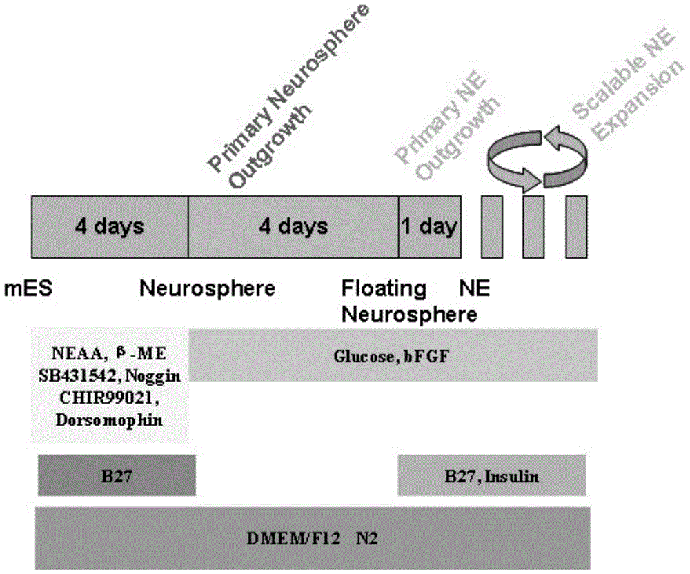
[0081] First, 24-well plates (Corning) were coated with Matrigel (BD). After cell counting, mouse embryonic stem cells (mESCs) were plated at 1x10 3 The number per well was seeded on a cell culture plate. Add 400 μl of culture medium to each well, and change the medium every other day. After 3-5 days, the cells grow into clump-like clones, and then start induction, which is divided into 3 stages.
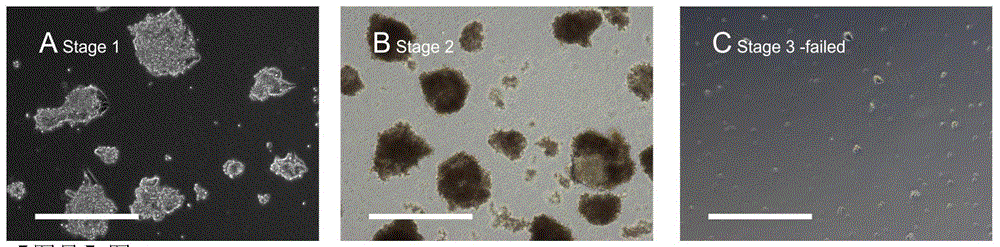
[0082] 1) The first stage: the medium of the adherent cultured mouse embryonic stem cell cluster clones was replaced with the fir...
Embodiment 2
[0089] Example 2 Immunological identification of mouse neuroepithelial cells
[0090] The first to third generation of neuroepithelial cells obtained in Example 1 were subjected to immunofluorescence detection, and the specific steps included fixation, membrane rupture, and blocking solution for blocking for 1 hour, and then adding primary antibodies at the ratio of Nestin (Beyotime) 1:10 , Sox2 (CST) 1:200, Sox1 (R&D) 1:10, Pax6 (Abcam) 1:20, CD133 (Bioss) 1:100 or Sox9 (Millipore) 1:5000 overnight at 4°C, after washing with PBS, add a Anti-appropriate fluorescent secondary antibody FITC (ImmunoResearch), CY3 (ImmunoResearch) was incubated at room temperature for 2 hours. Finally, after washing with PBS, the cells were stained with Hoechst or DAPI and mounted with a mounting agent. Detection was performed under a fluorescence microscope (Zeiss) and a scanning laser confocal imaging system (Leica). Immunofluorescent staining of NE revealed expression of the neural progenitor...
Embodiment 3
[0091] Example 3 Identification of polar structure of neuroepithelial cells and neural rosette-shaped specific markers The neuroepithelial cells obtained in Example 1 were subjected to cell immunofluorescence staining, the operation method was the same as in Example 2, and the primary antibodies used were respectively PLZF (Santa Cruz) 1:50, ZO-1 (Santa Cruz) 1:50, SOX2 (CST) 1:200, PAX6 (Abcam) 1:200, the results show that the neuroepithelial cells obtained in Example 1 show nuclear expression transcription Factors PLZF and DACH1, and the expression of neuroepithelial polarity marker ZO-1 was also found (such as Figure 6 As shown, A. NE-specific transcription factor DACH1, also expresses neural precursor marker Nestin. B. NE can also be stained by the neural precursor transcription factor Sox2. C.NE can express the polarity marker ZO-1, indicating the polarity characteristics of neuroepithelial cells, and can also express the neural precursor marker Pax6. D.NE expresses it...
PUM
 Abstract
Description
Claims
Application Information