Myoelectric Pain Measuring Device

A technology for measuring pain and electromyography, applied in the field of medical devices, can solve the problems of not easy to carry, inaccurate test results, relying on subjective pain reporting, etc., and achieves the effects of reliability and validity, reduced instrument size, and easy portability.

Active Publication Date: 2017-03-08
HUAZHONG UNIV OF SCI & TECH
View PDF4 Cites 0 Cited by
  • Summary
  • Abstract
  • Description
  • Claims
  • Application Information

AI Technical Summary

Problems solved by technology

[0006] In view of the above defects or improvement needs of the prior art, the present invention provides a myoelectric pain measurement device, the purpose of which is to remove noise through instruments and programs, replace the detection of RⅢ reflex, and measure the pain threshold, thereby solving the problem of existing clinical pain measurement. The method relies on subjective pain reporting, the test results are inaccurate, and the experimental equipment is large in scale and not easy to carry, etc.

Method used


Image

  • Myoelectric Pain Measuring Device
  • Myoelectric Pain Measuring Device

Examples


Embodiment 1

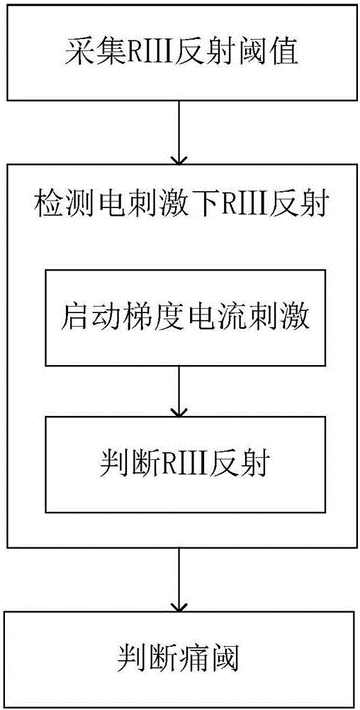
[0053] A kind of myoelectric pain measurement method, comprises the following steps:

[0054] (1) Acquisition of RⅢ reflex threshold: set a reference electrode at the biceps femoris tendon; set a working electrode at the abdomen of the short head of the biceps femoris, and both the reference electrode and the working electrode are patch electrodes; record the voltage The difference was taken as the background discharge of the biceps femoris, and 1.5 times of the background discharge was taken as the RⅢ reflex threshold.

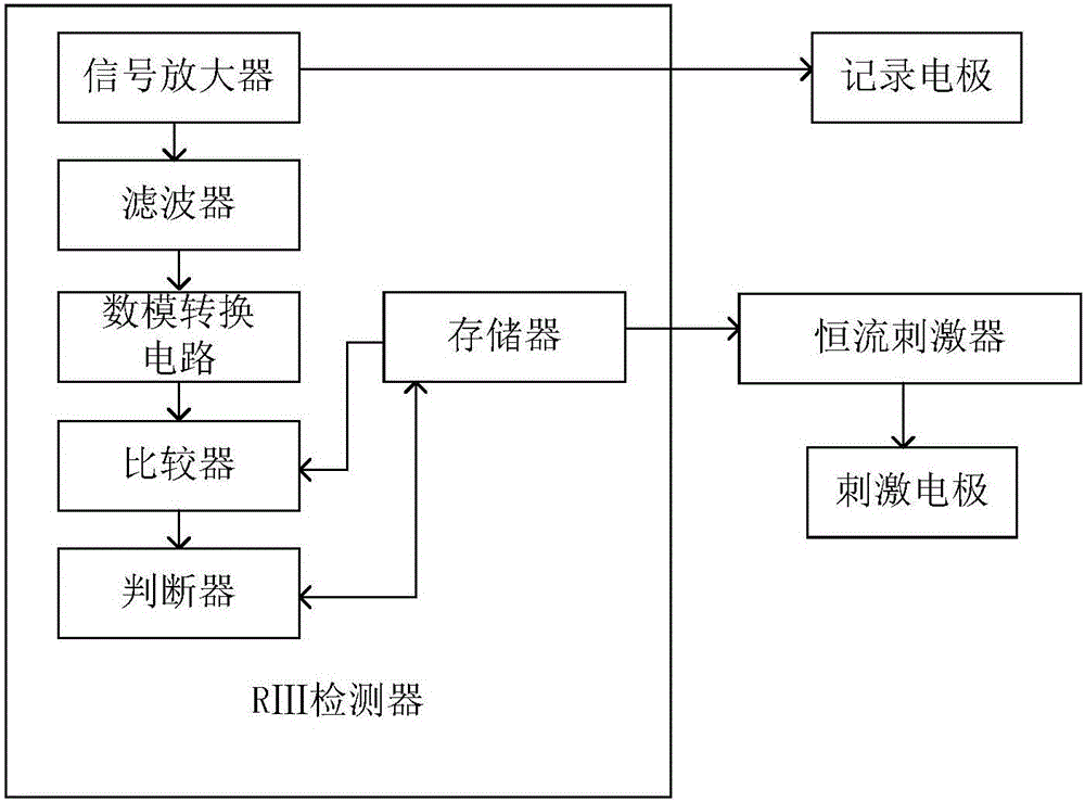
[0055] (2) Detection of RⅢ reflex under electrical stimulation:

[0056] (2-1) Start current gradient increasing stimulation, each gradient repeats an odd number of times 7, and the interval between two stimulations is 5 seconds, and each stimulation step is:

[0057] The peroneal nerve behind the lateral ankle joint is stimulated according to the gradient current value. The wave width of the current stimulation is 1ms and the frequency is 20Hz; the peak vol...

Embodiment 2

[0061] A kind of myoelectric pain measurement method, comprises the following steps:

[0062] (1) Acquisition of RⅢ reflex threshold: set a reference electrode at the biceps femoris tendon; set a working electrode at the abdomen of the short head of the biceps femoris, and both the reference electrode and the working electrode are patch electrodes; record the voltage The difference was taken as the background discharge of the biceps femoris, and twice the background discharge was taken as the RⅢ reflex threshold.

[0063] (2) Detection of RⅢ reflex under electrical stimulation:

[0064] (2-1) Start current gradient increasing stimulation, each gradient repeats an odd number of 5 times, and the interval between two stimulations is 10 seconds, and each stimulation step is:

[0065] The peroneal nerve behind the lateral ankle joint is stimulated according to the gradient current value. The wave width of the current stimulation is 5ms and the frequency is 10Hz; the peak voltage o...

Embodiment 3

[0069] A kind of myoelectric pain measurement method, comprises the following steps:

[0070] (1) Acquisition of RⅢ reflex threshold: set a reference electrode at the biceps femoris tendon; set a working electrode at the abdomen of the short head of the biceps femoris, and both the reference electrode and the working electrode are patch electrodes; record the voltage The difference was taken as the background discharge of the biceps femoris, and 2.2 times of the background discharge was taken as the RⅢ reflex threshold.

[0071] (2) Detection of RⅢ reflex under electrical stimulation:

[0072] (2-1) Start current gradient decreasing stimulation, repeat each gradient for an odd number of times 3, and the interval between two stimulations is 15 seconds. The steps of each stimulation are:

[0073] The peroneal nerve behind the lateral malleolus is stimulated according to the gradient current value. The wave width of the current stimulation is 10ms and the frequency is 0.1Hz; the...


PUM


Abstract

The invention discloses a myoelectricity pain measurement method and device for clinical use. The method comprises the steps of (1) acquiring an RIII reflex threshold, (2) detecting RIII reflex under electrical stimulation, and (3) judging the threshold of pain. The device comprises a stimulating electrode, a recording electrode, a constant-current stimulator and an RIII detector, wherein the stimulating electrode is electrically connected with the constant-current stimulator, the constant-current stimulator is in data connection with the RIII detector, the recording electrode is in data connection with the RIII detector, and the RIII detector comprises a signal amplifier, a filter, an analog-digital conversion circuit, a memory, a comparator and a judger and is used for recording the output current of the constant-current stimulator, recording the voltage data recorded by the recording electrode, and judging the threshold of pain according to the pain measurement method. The myoelectricity pain measurement method and device for clinical use solve the technical problem that according to an existing clinic pain measurement method, the measurement result is not accurate due to the fact that judgment is made according to subjective pain reporting.

Description

technical field [0001] The invention belongs to the field of medical devices, and more specifically relates to a myoelectric pain measuring device. Background technique [0002] Pain is a complex psychobiological process caused by noxious stimuli inside and outside the body. It is a common symptom of certain clinical diseases. It is not only a physiological feeling type, but also an unpleasant emotion in psychology. Pain is a subjective experience. Quantitative analysis of this subjective feeling is necessary for clinical work. It is directly related to the diagnosis and classification of patients, the selection of treatment methods, the observation of changes in the condition, the evaluation of treatment effects, and the research work on pain. However, the clinical measurement of pain largely depends on the oral dialogue between the patient and the doctor or nurse. There are no instruments and equipment that can quantitatively evaluate the pain intensity, independently and ...

Claims


Application Information

Patent Timeline
Patent Type & Authority Patents(China)
IPC IPC(8): A61B5/0488A61B5/00
CPCA61B5/4827
Inventor 李熳潘晓莉王俊俊刘双喜张力宋元龙郭乐江裴磊
Owner HUAZHONG UNIV OF SCI & TECH
Why Eureka
Try Eureka