Device and method for carrying out comparison and analysis according to different blood anticoagulation effects
A blood anticoagulant and comparative analysis technology, applied in the direction of transmittance measurement, etc., can solve problems such as poor definition
- Summary
- Abstract
- Description
- Claims
- Application Information
AI Technical Summary
Problems solved by technology
Method used
Image

Examples
Embodiment 1
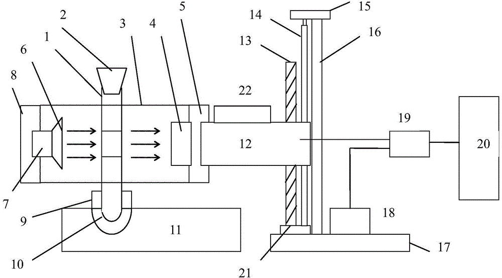
[0044] A device for comparative analysis of the effects of different blood anticoagulants, which includes a light source array, a detector array, a detection rack, a sample tube, a sample tube holder, a detection lifting system, a control module, and a PC;
[0045] The detection and lifting system consists of a horizontal bracket, a lifting screw, a vertical guide rail, a support rod, a supporting top seat, a lifting base, a lifting seat, and a stepping motor. The stepping motor is connected to the control module by communication, and the stepping motor also has a governor. , directly controlled by the PC through the control module;
[0046] The light source array is composed of multiple monochromatic light sources arranged in a row;
[0047] The detector array is composed of multiple detectors, and each monochromatic light source is directly opposite to a detector on a horizontal line across a sample tube;
[0048] There is also an optical path use module, which is controlle...
Embodiment 2
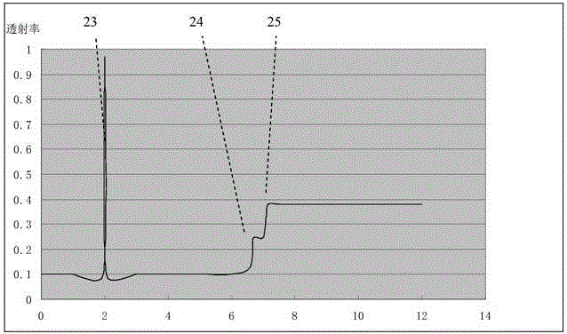
[0071] The difference between the implementation of Example 2 and Example 1 is that the wavelength of the monochromatic light source used is 670nm, and the four mixed anticoagulants added in the blood sample sources A and B are EDTA-K2, heparin sodium, citron Sodium oxalate, potassium oxalate, and sodium citrate are all mixed with an anticoagulant in a ratio of 1:4. The concentration of the anticoagulant is added to the general concentration of the anticoagulant. After 6 hours of transmittance scanning, step 3) can be carried out again. Scanning, and forming an image of the scanning data, and comparing with the image formed of the scanning data of the same sample 6 hours ago, to determine the change of the layer gap, thereby further determining the change of the anticoagulant performance. Except for the blood sample added with EDTA-K2 and potassium oxalate 1:4 anticoagulant, the boundary of blood component stratification was slightly blurred after 6 hours, and other changes wer...
Embodiment 3
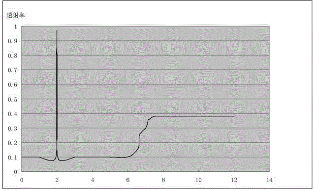
[0073]The embodiment of Example 3 is different from Example 2 in that the wavelength of the monochromatic light source used is 690 nm, and the mixed anticoagulants added in the blood sample sources A and B are EDTA-K2 and heparin sodium 1:4, 1:4, respectively. 6, and the anticoagulant mixed with EDTA-K2 and lithium heparin at a ratio of 1:5 and 1:9. The concentration of the anticoagulant is usually the concentration of the anticoagulant. Step 3 can be performed again 6 hours after the transmittance scan. ), and imaged the scan data and compared the image formed by the scan data of the same sample 6 hours earlier to determine the change in the delamination gap, thereby further determining the change in the anticoagulant performance. Through the comparison of the transmittance of the blood sample and the observation after magnification, it was found that the blood sample added with the anticoagulant mixed with EDTA-K2 and lithium heparin had a clearer blood sample stratification ...
PUM
 Abstract
Description
Claims
Application Information