Arteriovenous retinal vessel segmentation method for eye fundus image

A technology for retinal blood vessels and fundus images, applied in image analysis, image enhancement, image data processing, etc., can solve problems such as low degree of automation

Active Publication Date: 2015-04-22
杭州求是创新健康科技有限公司
View PDF6 Cites 14 Cited by
  • Summary
  • Abstract
  • Description
  • Claims
  • Application Information

AI Technical Summary

Problems solved by technology

[0007] Blood vessel segmentation, optic disc positioning and vessel classification (arteriovenous split) on fundus images are the basis of retinal v...

Method used


Image

  • Arteriovenous retinal vessel segmentation method for eye fundus image
  • Arteriovenous retinal vessel segmentation method for eye fundus image
  • Arteriovenous retinal vessel segmentation method for eye fundus image

Examples


Embodiment Construction

[0033] The present invention will be described in detail below with reference to the drawings and specific embodiments.

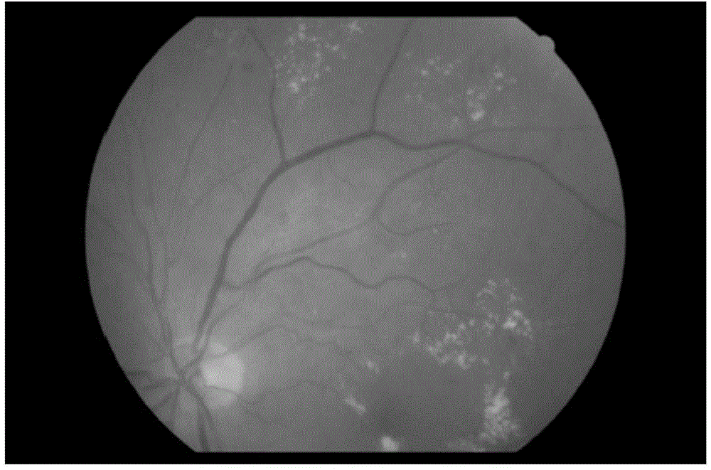
[0034] This embodiment takes figure 1 The fundus image shown is taken as an example to illustrate the arteriovenous retinal vessel segmentation method of the fundus image, and the size of the fundus image is 3000×3000. There are bright rings in the fundus image due to the ring reflection caused by photography, the non-vascular step edge around the optic disc, patchy lesions, and hemorrhagic lesions.

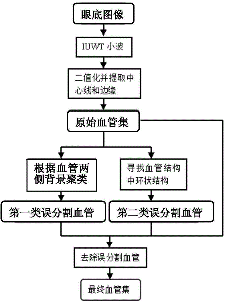
[0035] The classification process for the arteriovenous retinal vessel segmentation of the fundus image is as follows: figure 2 shown, including the following steps:

[0036] (1-1) Carry out wavelet transform (IUWT wavelet) to fundus image, carry out binarization process to the fundus image after wavelet transform according to preset binarization threshold value, and extract the center in the fundus image after binarization process lines and edges, to get ...


PUM


Abstract

The invention discloses an arteriovenous retinal vessel segmentation method for an eye fundus image. The method comprises the following steps that binarization processing is carried out on a pre-processed eye fundus image according to a preset image-binary threshold value, the central line and the edge in the eye fundus image after the binarization processing are extracted, and a blood vessel tree is obtained; disconnection process is carried out on the branching portions of the blood vessel tree to obtain blood vessel segments, line segmentation is carried out on all the blood vessel segments to obtain blood vessels, and an original blood vessel set is obtained; mis-segmented blood vessels are determined and removed from the original blood vessel set to obtain a global blood vessel set. According to the method, after the original blood vessel set is obtained, the mis-segmented blood vessels are further determined through the background and the shapes of the blood vessels, mis-segmented blood vessels which are caused by annular light reflection of photograph, non-blood-vessel step edges around an optic disc, porphyritic lesions, bleeding lesions and other reasons can be removed effectively, and the segmentation precision of the blood vessels is improved.

Description

technical field [0001] The invention relates to the technical field of computer-aided diagnosis, in particular to a method for segmenting arteriovenous retinal blood vessels of fundus images. Background technique [0002] With the rapid development of the field of artificial intelligence in computer technology, computer-aided diagnosis technology is also gradually developed. Computer-aided diagnosis technology refers to the use of imaging, medical image processing technology and other possible physiological and biochemical means, combined with computer analysis and calculation, to assist radiologists in finding lesions and improving the accuracy of diagnosis. [0003] Usually, computer-aided diagnosis in medical imaging is divided into three steps, as follows: the first step is to extract the lesion from the normal structure; the second step is to quantify the image features; the third step is to process the data and draw conclusions . [0004] Because the computer can ful...

Claims


Application Information

Patent Timeline
IPC IPC(8): G06T7/00
CPCG06T2207/30041G06T2207/30101
Inventor 吴健黎罗河邓水光李莹尹建伟吴朝晖
Owner 杭州求是创新健康科技有限公司
Why Eureka
Try Eureka