
Adjacent dual-X-ray source CT imaging system and application thereof

A CT imaging and system application technology, applied in computed tomography scanners, echo tomography, etc., can solve problems such as eliminating motion artifacts, increasing cardiac imaging radiation dose, and ineffective use of X-ray projection data.

Inactive Publication Date: 2015-04-29
SHENZHEN INST OF ADVANCED TECH CHINESE ACAD OF SCI
View PDF5 Cites 7 Cited by
  • Summary
  • Abstract
  • Description
  • Claims
  • Application Information

AI Technical Summary

Problems solved by technology

The problem with this gating technique is that some of the X-ray projection data is not used effectively, greatly increasing the radiation dose for cardiac imaging
Therefore, it is impossible to completely eliminate motion artifacts simply by increasing the scanning speed

Method used

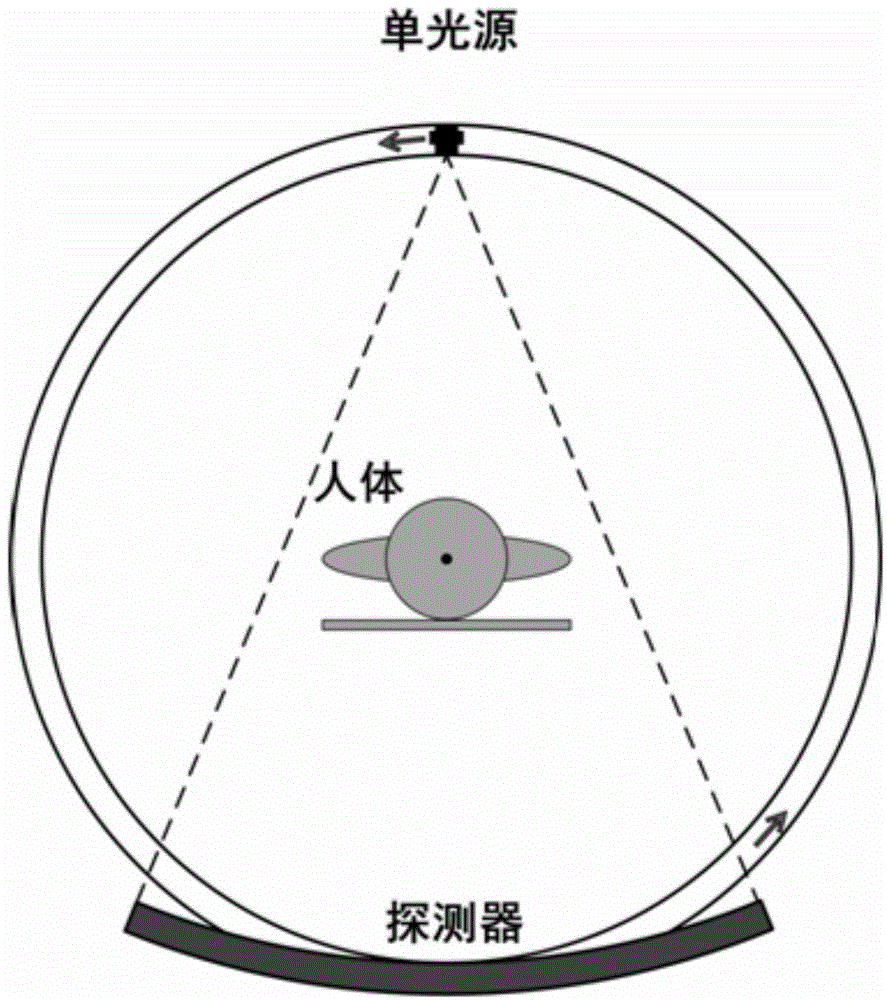
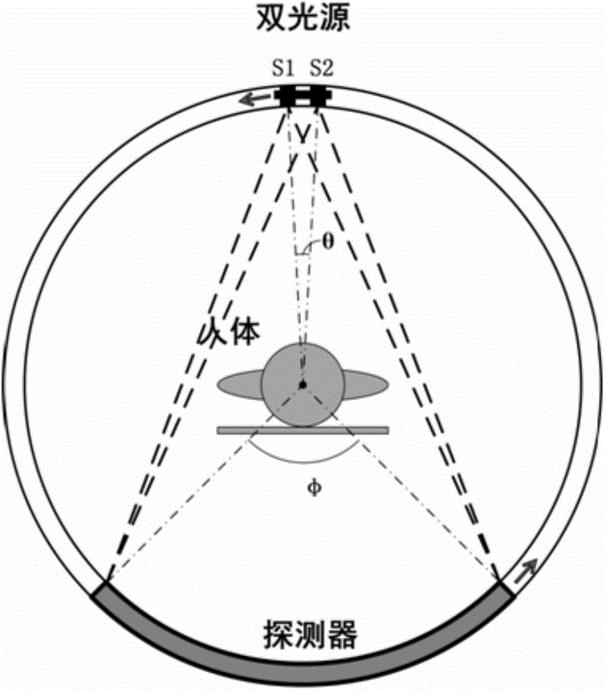
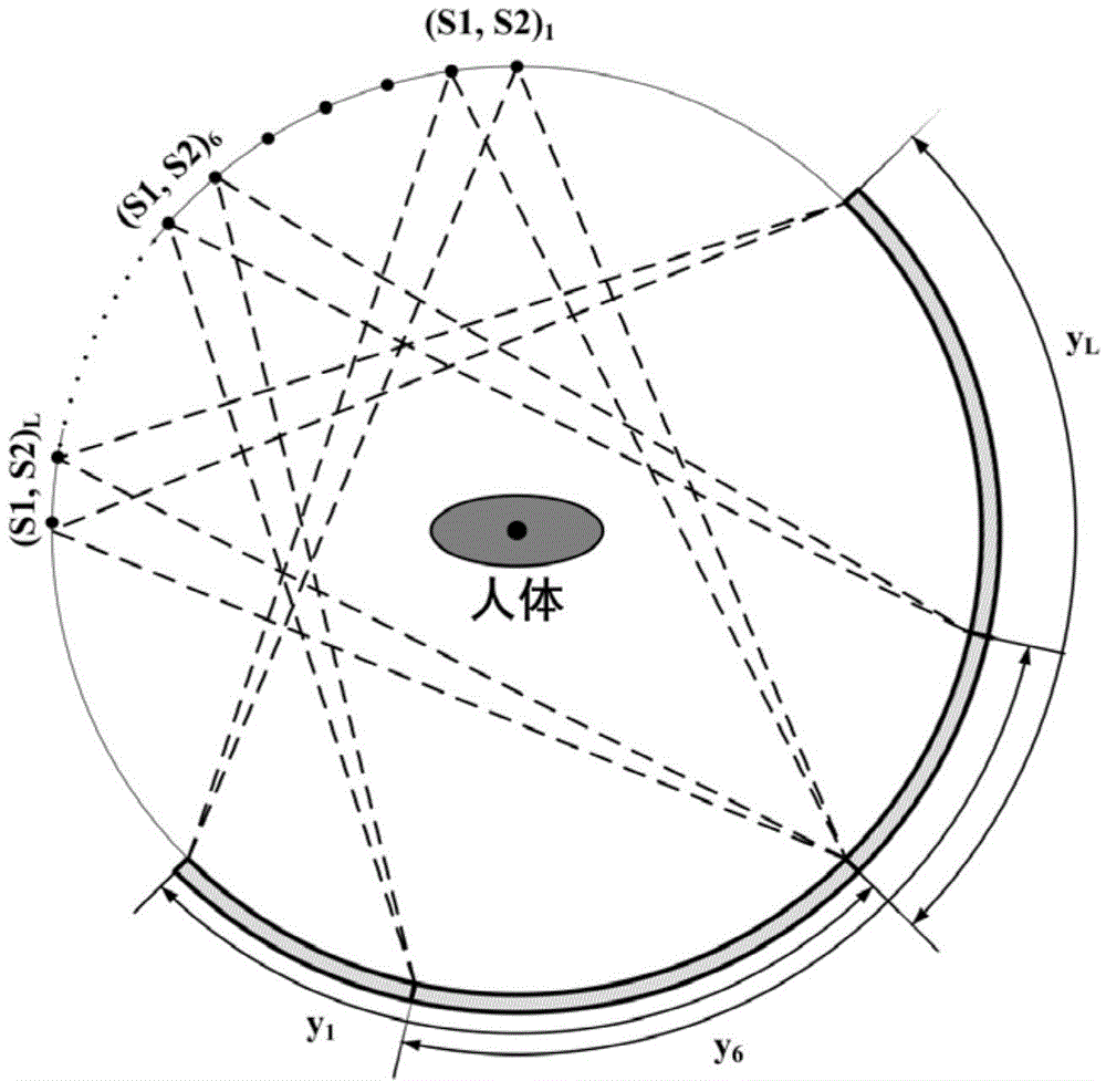

Image

  • Adjacent dual-X-ray source CT imaging system and application thereof
  • Adjacent dual-X-ray source CT imaging system and application thereof
  • Adjacent dual-X-ray source CT imaging system and application thereof

Examples


Embodiment 1

[0047] Such as figure 2 As shown, an adjacent double X-ray source CT imaging system includes an X light source and a detector; the X light sources are adjacent double X light sources S1 and S2, and the adjacent double X light sources are directly opposite to the detector set up.

[0048] The photon doses emitted by the adjacent double X light sources are the same.

[0049] The adjacent double X light sources are parallel to the rotation plane.

[0050] The X light source is an X-ray light source;

[0051] The center of the detector is located at the center of rotation;

[0052] The rotation center refers to the center where adjacent double X light sources and detectors rotate synchronously.

[0053] The detector is an arc detector.

[0054] A separation and reconstruction algorithm for processing projection images obtained by the above-mentioned adjacent dual X-ray source CT imaging system comprises the following steps:

[0055] (1) Assume that the Nth projection under ...


PUM


Abstract

The invention belongs to the technical field of CT imaging systems, and particularly discloses an adjacent dual X-ray source CT imaging system and application thereof. The adjacent dual X-ray source CT imaging system comprises X light sources and a detector; the X light sources are adjacent dual X light sources; and the adjacent dual X light sources and the detector are oppositely arranged. The doses of photons emitted by the adjacent dual X light sources are the same. The adjacent dual X light sources are parallel to a rotation panel. According to the adjacent dual X-ray source CT imaging system, the adjacent dual X light sources work at the same time; on one hand, the operating power of the light sources can be reduced, so that the service life of a ray tube is accordingly prolonged; on the other hand, motion artifact of images can be effectively reduced. The adjacent dual X-ray source CT imaging system is low in implementation cost; and large structure improvements on an existing CT device are not needed. The adjacent dual X-ray source CT imaging system can be applied to the medical treatment CT or the industrial CT with the large radiation exposure dose.

Description

【Technical field】 [0001] The invention belongs to the technical field of CT imaging, and in particular relates to a CT imaging system with adjacent double X-ray sources and its application. 【Background technique】 [0002] Since Hounsfield invented the first CT machine in 1972, CT technology has had a revolutionary impact on medical diagnosis and industrial non-destructive testing. CT has become one of the important detection methods in medical, biological, aerospace, defense and other industries. [0003] In 2005, Siemens launched the world's first dual-source CT (DSCT) system - SOMATOM Definition at the Radiology Congress of North America (RSNA). Based on the mature SOMATOM Sensation64 technology and Straton zero mega metal tube, Two sets of 64-layer image data acquisition systems are integrated in the frame so that a high-quality image can be obtained after the entire frame is rotated by 90°. It takes 0.33s for the gantry to rotate a circle, but the image acquisition can ...

Claims


Application Information

Patent Timeline
IPC IPC(8): A61B6/03
Inventor 安谋张翠李荣茂谢耀钦
Owner SHENZHEN INST OF ADVANCED TECH CHINESE ACAD OF SCI
Learn More