A computerized automatic grading method for degeneration of intervertebral disc images

A degenerative and automatic grading technology, applied in the field of clinical medicine, can solve the problems that doctors are prone to errors in grading degenerative changes, and there is no quantitative grading standard.

Inactive Publication Date: 2018-03-20
THE THIRD AFFILIATED HOSPITAL OF THIRD MILITARY MEDICAL UNIV OF PLA
View PDF1 Cites 0 Cited by
  • Summary
  • Abstract
  • Description
  • Claims
  • Application Information

AI Technical Summary

Problems solved by technology

[0004] The above-mentioned methods only realize the localization of the intervertebral disc or only the localization of the normal intervertebral disc, but there is no quantitative grading standard for the degree of lesion
In clinical practice, due to the lack of quantitative assessment standards, doctors are prone to errors in grading degeneration
Judgment of intervertebral disc herniation based on the grayscale and texture information of the intervertebral disc, but there is no relevant literature on the grading of degeneration

Method used


Image

  • A computerized automatic grading method for degeneration of intervertebral disc images
  • A computerized automatic grading method for degeneration of intervertebral disc images
  • A computerized automatic grading method for degeneration of intervertebral disc images

Examples


Embodiment 1

[0050] As shown in the figure, the degeneration computer automatic grading method of a kind of intervertebral disc image provided by the present invention comprises the following steps:

[0051] S1: Obtain an image of the intervertebral disc, and obtain a sample image through image preprocessing;

[0052] S2: Extract the intervertebral disc position from the intervertebral disc image to obtain an oval intervertebral disc area;

[0053] S3: Calculating the gray ratio of the nucleus pulposus and the annulus fibrosus in the oval intervertebral disc region;

[0054] S4: Grading the degeneration of the intervertebral disc image according to the gray ratio.

[0055] The oval intervertebral disc area is formed in the following steps:

[0056] S21: Obtain the rectangular area of ​​each intervertebral disc through a positioning algorithm;

[0057] S22: Process the intervertebral disc image with a Gabor filter to obtain filter images of different frequencies;

[0058] S23: Calculate...

Embodiment 2

[0081] The difference between this embodiment and embodiment 1 is only:

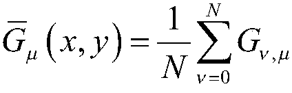
[0082] figure 2 In (2a) segmented intervertebral disc region based on positioning results; (2b) intervertebral disc boundary determined by Gabor features; (2c) edge point detection; (2d) ellipse fitting; image 3 (3a) Negative values ​​in the Gabor coefficient graph are set to zero; (3b) G(n); (3c) spine extraction results; Figure 4 (4a) intervertebral disc characteristics; (4b) G h (n) Curve; (4c) Intervertebral disc boundary determined by Gabor features; (4d) Comparison of positioning results before and after algorithm improvement; Figure 8 (8a) Level 2; (8b) Level 3; (8c) Level 4; (8d) Level 5;

[0083] The Gabor wavelet transform used in this embodiment is a windowed Fourier transform; in the spatial domain, the impulse response of the two-dimensional Gabor filter is the result obtained by multiplying the complex exponential oscillatory function by the Gaussian envelope function:

[0084]

...


PUM


Abstract

The invention discloses an automatic computer grading method for retrogression of intervertebral disc images. The method comprises the steps that edge information of a spine and intervertebral discs is obtained by processing a series of spine Gabor characteristic images; a spine area is extracted based on the edge information of the spine; the intervertebral discs are located in the spine area according to the information and prior information of positions of the intervertebral discs; nucleus pulposus and fibrous ring grey scale information is obtained by combining an ellipse fitting method according to a locating result and a Gabor coefficient diagram; and finally grading is realized according to the grey scale information and geometrical information. The method effectively reduces a target search range by utilizing the multi-scale and multi-direction characteristic of a Gabor wavelet and combining direction information, realizes full-automatic and unsupervised recognition and grading by extracting texture information of the intervertebral discs on a sagittal diagram via the Gabor wavelet, and is applicable to the pathological and non-pathological intervertebral discs. An accuracy rate and the feasibility of an algorithm are verified via MRI (Magnetic Resonance Imaging) data of 37 patients, and grading evaluation of the retrogression in first 5 grades is realized on the basis that the locating accuracy rate can reach 96.6%.

Description

technical field [0001] The invention relates to the technical field of clinical medicine, in particular to a computer automatic grading method for degeneration of intervertebral disc images. Background technique [0002] Magnetic resonance imaging can produce detailed structural features and clear images of internal human organs because of the inherent versatility of this modality, which is amenable to non-invasive medical evaluation of tissue anatomy, pathology, metabolism, and flow. In addition, compared with other forms of images, MRI images have significant advantages such as high spatial resolution, flexible scanning angles, and no bone artifacts. Therefore, with the increasing demand for computer-assisted therapy in clinical applications, the computer analysis of magnetic resonance (Magnetic Resonance, MR) spine images has become more and more important for spine diagnosis. [0003] For the positioning of MRI intervertebral discs, from the beginning, only the thoracic...

Claims


Application Information

Patent Timeline
Patent Type & Authority Patents(China)
IPC IPC(8): G06T7/00G06K9/62A61B5/055
CPCA61B5/055G06T7/0012G06T2207/20064G06T2207/30012
Inventor 朱新建林晓红闫铮冯正权闫庆广何庆华
Owner THE THIRD AFFILIATED HOSPITAL OF THIRD MILITARY MEDICAL UNIV OF PLA
Why Eureka
Try Eureka