Ultrasonographic perfusion parameter imaging method based on single-pixel TIC source

A technology of contrast-enhanced ultrasound and imaging methods, which is applied in ultrasonic/sonic/infrasonic diagnosis, sonic diagnosis, infrasonic diagnosis, etc. It can solve problems such as subjective differences, difficulty in repeated implementation, and lack of detailed information, so as to improve SCR and suppress clutter The effect of interference and rich perfusion detail information

Active Publication Date: 2015-06-10
XI AN JIAOTONG UNIV
View PDF5 Cites 9 Cited by
  • Summary
  • Abstract
  • Description
  • Claims
  • Application Information

AI Technical Summary

Problems solved by technology

[0005] Aiming at the limitations of extraction of TIC under conventional large ROI for ultrasound-enhanced blood perfusion parameter imaging (subjective variability, difficulty in repeated implementation, lack o

Method used


Image

  • Ultrasonographic perfusion parameter imaging method based on single-pixel TIC source
  • Ultrasonographic perfusion parameter imaging method based on single-pixel TIC source
  • Ultrasonographic perfusion parameter imaging method based on single-pixel TIC source

Examples


Embodiment Construction

[0035] The present invention will be described in detail below in conjunction with the accompanying drawings and embodiments.

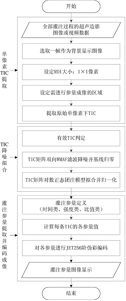
[0036] The present invention mainly proposes to extract TIC under the minimum unit ROI, ie "single pixel", to perform ultrasound-enhanced blood flow perfusion parameter imaging under group perfusion, and its core is to suppress the strong clutter interference of TIC under single pixel. For this reason, the present invention proposes the preprocessing scheme to TIC: (1) at first all TICs that are extracted under the single pixel of the imaging area to be treated are subjected to correlation-based validity identification, to eliminate invalid TICs, and save calculation time; (2) ) Secondly, the two-way weighted moving average filter (WMAF) is used to suppress the interference of clutter on TIC and avoid the interference of time shift; (3) Finally, the bolus perfusion model is used to fit the contrast microbubbles. Accurately obtain high SCR TIC while ci...


PUM


Abstract

The invention provides an ultrasonographic perfusion parameter imaging method based on a single-pixel TIC source. The method comprises the following steps: extracting original TIC under each pixel unit of an ultrasonographic sequence video image; carrying out effective TIC screening, bidirectional weighted moving average filtering and bolus perfusion model fitting on the original TIC to obtain a high-SCR single-pixel TIC matrix; respectively extracting time class, strength class and specific value class of perfusion parameter matrixes in the single-pixel TIC matrix, and carrying out pseudo-color encoding. The process of extracting the TIC of the ultrasonographic sequence video image by single pixel ROI is automatically carried out, and the SCR of the TIC under the single-pixel ROI is greatly improved and the interference of noise wave is inhibited by virtue of the combination of bidirectional weighted moving average filtering and lognormal bolus perfusion model fitting. Compared with traditional perfusion parameter evaluation and imaging, the ultrasonic blood perfusion parameter imaging based on the single-pixel TIC source contains the most abundant detail information and is more visual.

Description

technical field [0001] The invention relates to the technical field of contrast-enhanced ultrasound imaging, in particular to an imaging method of ultrasound blood perfusion parameters based on a single-pixel TIC source. Background technique [0002] The development of contrast-enhanced ultrasound microbubbles and contrast-enhanced imaging methods has made it possible to quantitatively estimate blood perfusion with contrast-enhanced ultrasound. Although the spatial resolution of contrast-enhanced ultrasound imaging is limited and individual capillaries cannot be identified, the echo signal intensity caused by contrast-enhanced microbubbles is proportional to the concentration of microbubbles and also proportional to tissue blood volume. The discovery of this proportional relationship has been made in recent years To promote the development of an important application of contrast-enhanced ultrasound imaging, that is, the estimation of perfusion parameters. Contrast-enhanced ...

Claims


Application Information

Patent Timeline
IPC IPC(8): A61B8/06
CPCA61B8/06A61B8/481A61B8/5207A61B8/5292
Inventor 万明习王弟亚钟徽杨萱韩萌肖梦楠
Owner XI AN JIAOTONG UNIV
Why Eureka
Try Eureka