Kit for thoracolumbar vertebral pedicle operation, and use method thereof

A pedicle and surgery technology, applied in the medical field, can solve the problems of large radiation damage to patients and physicians, difficulty in finding the pedicle area, and difficulty in accurate success of the skin, so as to shorten the operation time, reduce tissue damage and bleeding , the effect of reducing the risk of X-ray radiation

Inactive Publication Date: 2015-08-26
黄象望 +1
View PDF4 Cites 0 Cited by
  • Summary
  • Abstract
  • Description
  • Claims
  • Application Information

AI Technical Summary

Problems solved by technology

In the current operation, an extracutaneous grid locator is often used to locate the lesion, and the adhesive tape is fixed on the skin at the position of the injured vertebra. For the first time, fluoroscopy is performed to analyze and accurately locate the position of the pedicle interval of the injured vertebra under the grid image. , traced on the skin, use a single needle or directly use the pedicle drill at this position, because it is difficult to find the pedicle interval (2° to the right and 10° to the left) due to the sliding of the skin, and repeated puncture and fluoroscopy
The simple Kirschner wire positioning or grid positioning adopted in the prior art is difficult to succeed precisely because the swinging skin of the patient's body position can be moved; the percutaneous pedicle drill is used for soft tissue (skin, subcutaneous, fascia and back muscles) ) to find the pedicle interval (2° to the right and 10° to the left), which requires repeated punctures and repeated X-ray fluoroscopy, which takes a long time and causes great radiation damage to patients and doctors

Method used


Image

  • Kit for thoracolumbar vertebral pedicle operation, and use method thereof
  • Kit for thoracolumbar vertebral pedicle operation, and use method thereof
  • Kit for thoracolumbar vertebral pedicle operation, and use method thereof

Examples


Embodiment Construction

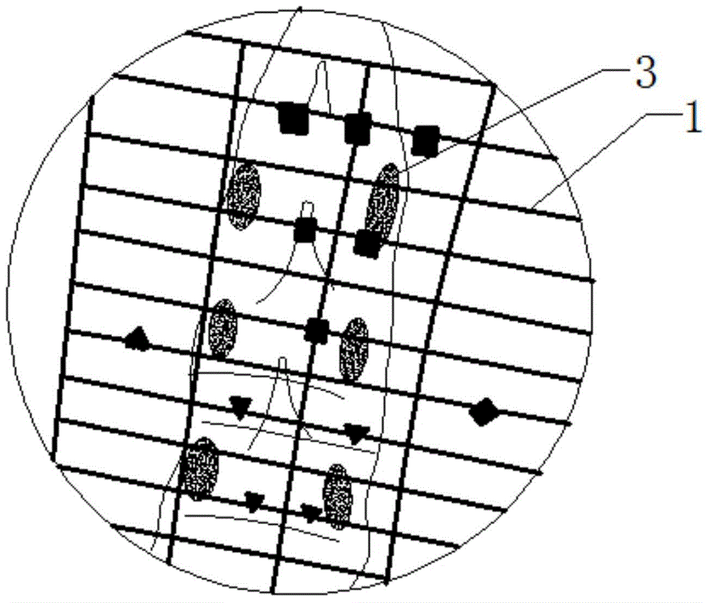
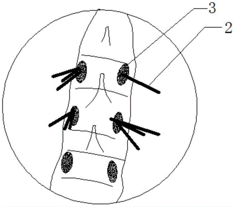
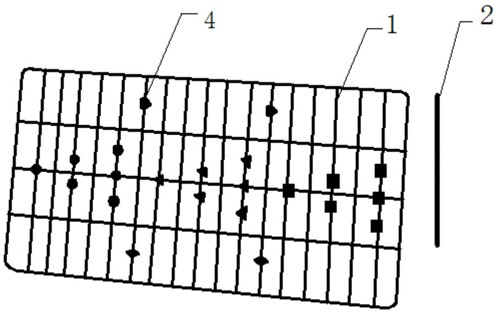
[0026] Such as Figure 1 to Figure 3 A thoracic and lumbar pedicle surgery kit as shown, including a developing grid 1, a Kirschner wire group and a perspective instrument (not shown); the developing grid 1 is closely attached to the skin of the lesion area of ​​the human body; Each K-wire set was inserted into 3 regions of each pedicle.

[0027] The Kirschner wire group is composed of three 2mm thick Kirschner wires 2 .

[0028] The fluoroscope (not shown) is X-ray fluoroscope, CT fluoroscopy or nuclear magnetic resonance imaging.

[0029] The developing grid is provided with a plurality of mark positions 4; it can well assist positioning.

[0030] A method of using a kit for thoracolumbar pedicle surgery, said method comprising the steps of:

[0031] Step 1: Put the patient prone on the see-through spinal support, and place his upper and lower limbs in a comfortable position;

[0032] Step 2: Determine the location of the patient's painful area;

[0033] Step 3: placing...


PUM


Abstract

The invention discloses a kit for thoracolumbar vertebral pedicle operation, and a use method thereof. The kit comprises a development grid, Kirschner wire sets and a scenograph, wherein the development grid is clung to the skin in the focus area; each Kirschner wire set is inserted into each vertebral pedicle area. The use method comprises the following steps: firstly, after perspective positioning of the development grid, the position of the injured vertebral pedicle is checked, and a puncture mark is made on the skin of the focus spine of a patient for accurate positioning; three Kirschner wires are adopted for percutaneous penetration in a triangular manner in each vertebral pedicle area, according to the vertebral pedicle areas (2 degrees on right and 10 degrees on left) after perspective treatment, the Kirschner wires at the optimal positions are indwelled, and screw paths are expanded to enter the vertebral body through the vertebral pedicle. According to the kit for thoracolumbar vertebral pedicle operation and the use method thereof, the development grid and the Kirschner wire sets are combined to realize successful one-time puncture positioning, so that repeated puncture is avoided, and the operation time is shortened; tissue damages and bleeding are reduced; the X-ray radiation risk of patients and physicians is reduced.

Description

technical field [0001] The invention relates to a set for thoracolumbar pedicle surgery and a use method thereof, belonging to the field of medical technology. Background technique [0002] In thoracolumbar pedicle surgery, minimally invasive percutaneous pedicle internal fixation is used in order to stabilize the spine, enhance the rigidity of the spine, prevent fractures and dislocations, and cause chronic pain in the spine. In the current operation, an extracutaneous grid locator is often used to locate the lesion, and the adhesive tape is fixed on the skin at the position of the injured vertebra. For the first time, fluoroscopy is performed to analyze and accurately locate the position of the pedicle of the injured vertebra under the grid image. , traced on the skin, use a single needle or directly use the pedicle drill at this position, because it is difficult to find the pedicle interval (2° to the right and 10° to the left) due to the sliding of the skin, and repeated...

Claims


Application Information

Patent Timeline
Patent Type & Authority Applications(China)
IPC IPC(8): A61B17/70A61B17/92
Inventor 黄象望刘宏哲
Owner 黄象望
Why Eureka
Try Eureka