Head-mounted stereo-display microsurgery operation system

A microsurgery and surgical system technology, applied in the field of medical equipment, can solve the problems of reducing the labor intensity of the operator, improving the operation efficiency of the operator, and being unable to put it into practical use, so as to reduce the physical labor intensity, have a large operating space, and be easy to use. The effect of implementation

Inactive Publication Date: 2015-11-25
顾雍舟
View PDF4 Cites 3 Cited by
  • Summary
  • Abstract
  • Description
  • Claims
  • Application Information

AI Technical Summary

Problems solved by technology

After patent retrieval, the author Zhu Cheng proposed a head-mounted wireless receiving virtual display surgical teaching system in the patent application number 2014101801100 (publication number CN103929625A) of "head-mounted wireless receiving virtual display surgical teaching system". This system is available For all surgical teaching, the idea is novel, but it cannot be used to improve the efficiency of surgery and reduce the labor intensity of the operator. The application of wireless communication is against the management of the operating room and interferes with other medical devices, so it cannot be put into practical use.

Method used


Image

  • Head-mounted stereo-display microsurgery operation system
  • Head-mounted stereo-display microsurgery operation system

Examples


specific Embodiment 1

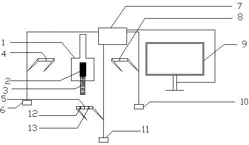
[0014] This embodiment utilizes a single-person binocular electronically controlled stereoscopic surgical microscope, and two high-definition cameras are installed at the eyepieces of the microscope. The operator wears a head-mounted stereoscopic image display, and a pair of miniature camera. The image signal captured by the high-definition camera is transmitted to the image processor, and the image processor converts the image captured by the high-definition camera into a normal image, a 180-degree inverted image, and a 90-degree rotated image, and sends them to the chief surgeon, the first assistant and the second surgeon respectively. Help the worn head-mounted stereoscopic image display to form a virtual stereoscopic image, and at the same time transmit the image to a large-screen display.

[0015] The miniature camera captures images in front of the operator. The head-mounted stereoscopic image display receives two channels of image signals, one from the image processor ...

specific Embodiment 2

[0016] This embodiment is a simple embodiment of the present invention. In this embodiment, a single-person monocular electronically controlled surgical microscope is used. A high-definition camera is installed at the eyepiece of the microscope, and the operator wears a head-mounted monocular image display. The image signal captured by the high-definition camera is transmitted to the image processor, and the image processor converts the image into an upright image, a 180-degree inverted image and a 90-degree rotated image, and sends them to the surgical head, the first surgical assistant and the second surgical assistant respectively. The head-mounted monocular image display forms a magnified image in front of the operator's eyes, and at the same time transmits the image to the large-screen display. The operator's chief surgeon, first assistant and second assistant can wear the head-mounted monocular image display, and the images they watch are just in line with the positive im...


PUM


Abstract

The invention relates to a head-mounted stereo-display microsurgery operation system. The head-mounted stereo-display microsurgery operation system comprises an operation stereoscopic microscope system, a high-definition camera, an image processor, head-mounted stereo-display systems, a display, minitype cameras and image conversion switches. The high-definition camera is used for collecting operation field images through the operation stereoscopic microscope system; the collected images are converted by the image processor according to the position of an operator; erect images, which each operator can see, are displayed on the head-mounted stereo-display system of each operator; the operators can observe an operation region and the environment of an operating room by utilizing the image conversion switches and the head-mounted minitype cameras. The head-mounted stereo-display microsurgery operation system has the advantages that the structure is simple, the use is convenient, the effect is remarkable, the operability is high and the implementation is easy.

Description

technical field [0001] The invention relates to a medical device, in particular to a head-mounted stereoscopic display microsurgical system. Background technique [0002] At present, all microsurgical operations rely on the operator to gaze at the surgical field of view through a fixed operating microscope to complete the operation. The operator needs to maintain a certain posture for a long time, which consumes a lot of energy, and the eyes are prone to fatigue. During the operation, intermittent rest is required to complete the operation. The distance between the microscope and the operation area is small, the operation space is narrow, and during the operation, because the observer or instructor cannot see the intraoperative image, it is impossible to understand and guide the operation. [0003] After searching the literature of the prior art, although the existing microsurgery system has a camera system, the images taken are not for the operator to watch, but are only us...

Claims


Application Information

Patent Timeline
Patent Type & Authority Applications(China)
IPC IPC(8): H04N7/18
Inventor 不公告发明人
Owner 顾雍舟
Why Eureka
Try Eureka