Method for detecting cavitation and ultrasonic medical equipment

A kind of medical equipment and ultrasound technology, applied in ultrasound therapy, ultrasound/sonic wave/infrasonic wave diagnosis, treatment, etc., can solve problems such as inaccurate detection, achieve the effect of preventing damage and shortening time

Active Publication Date: 2017-09-22
ALPINION MEDICAL SYST
View PDF1 Cites 0 Cited by
  • Summary
  • Abstract
  • Description
  • Claims
  • Application Information

AI Technical Summary

Problems solved by technology

However, the position where such cavitation occurs cannot be accurately detected, so damage to living tissue at the site of ultrasound treatment cannot be avoided

Method used


Image

  • Method for detecting cavitation and ultrasonic medical equipment
  • Method for detecting cavitation and ultrasonic medical equipment
  • Method for detecting cavitation and ultrasonic medical equipment

Examples


Embodiment Construction

[0020] Hereinafter, embodiments of the present invention will be described in detail with reference to the accompanying drawings.

[0021] The diagnostic images involved in the embodiments of the present invention may include B-mode images, C-mode images, and the like. Wherein, the β-mode image refers to the image mode showing the activity of the object to be measured, that is, the grayscale image. C-mode images refer to the color flow image mode. In addition, BC-mode image (BC-Mode Image) refers to an image mode that uses Doppler Effect (Doppler Effect) to display blood flow or body activity, as a mode that simultaneously provides β-mode image and C-mode image, An image modality that provides anatomical information along with blood flow and body activity information. That is, the B-mode is a grayscale image, which refers to the image mode showing the activity of the subject, and the C-mode is a color blood flow image, which refers to the image mode showing the blood flow or...


PUM


Abstract

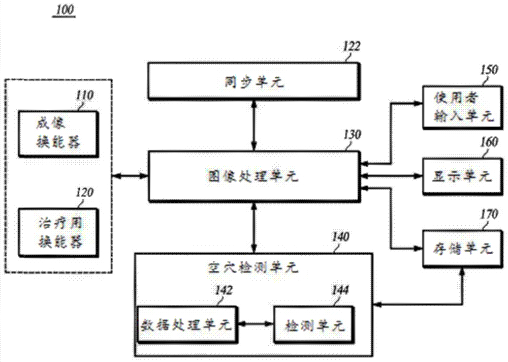
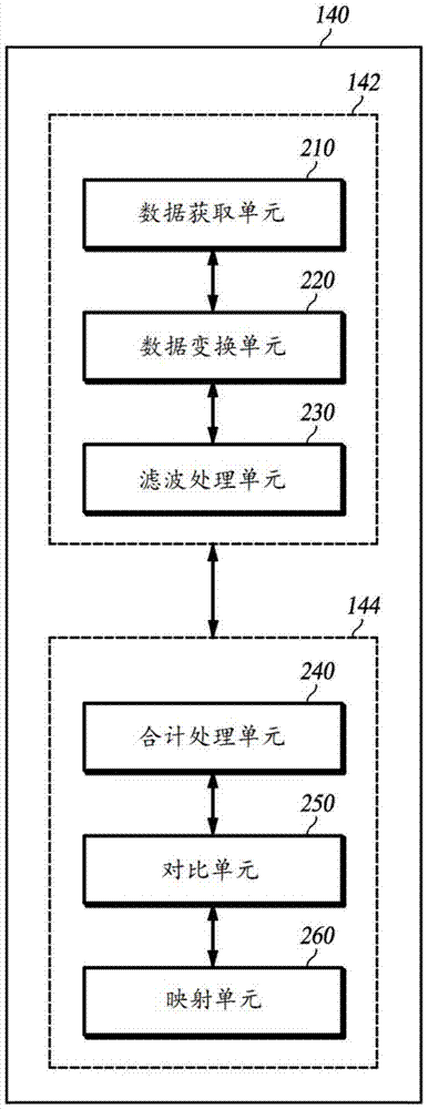
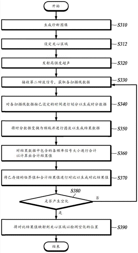
The invention discloses a method for detecting cavitation and ultrasonic medical equipment. The method for detecting cavitation and the ultrasonic medical equipment provided by the present invention process the reflection signal generated by focusing the ultrasound for imaging and high-intensity ultrasound on the object to be tested (the affected part of the patient). When cavitation is confirmed, the reflected The signal is subjected to data processing to detect the location of cavitation.

Description

technical field [0001] The invention relates to a method for detecting cavitation and ultrasonic medical equipment. More specifically, the present invention relates to a method and ultrasonic medical equipment for detecting the position of cavitation by processing reflected signals generated by focusing ultrasound for imaging and high-intensity ultrasound on a subject (patient's affected part). Background technique [0002] The description in this section is only to provide background technical information for the present invention, not to realize the prior art. [0003] Generally, High-Intensity Focused Ultrasound (HIFU: High-Intensity Focused Ultrasound) is used for the treatment of living tissues such as cancers, tumors, and lesions. That is, the high-intensity ultrasound treatment method is a method in which high-intensity ultrasound is emitted concentratedly at one point, and the thermal effect generated is used to cause necrosis of the target living tissue. It should...

Claims


Application Information

Patent Timeline
Patent Type & Authority Patents(China)
IPC IPC(8): A61B8/14A61N7/00
CPCA61B8/14A61B8/469A61B8/5207A61B8/5246A61N7/02A61N2007/0039A61B8/00
Inventor 孙健豪姜国珍金大昇全皙焕
Owner ALPINION MEDICAL SYST
Why Eureka
Try Eureka