Ultrasonic imaging method and device and ultrasonic equipment thereof

An ultrasonic imaging method and ultrasonic image technology, which are applied in ultrasonic/sonic/infrasonic diagnosis, sonic diagnosis, infrasonic diagnosis, etc., can solve the problems of time-consuming, large individual differences, proximity, etc. The effect of simplifying operating procedures

Active Publication Date: 2016-03-30
SONOSCAPE MEDICAL CORP
View PDF4 Cites 14 Cited by
  • Summary
  • Abstract
  • Description
  • Claims
  • Application Information

AI Technical Summary

Problems solved by technology

[0002] The traditional three-dimensional image observation of the endometrium is obtained by the doctor by manually adjusting the image. Due to the large individual differences, especially the endometrium of postpartum women has not fully recovered, the line echo of the end

Method used


Image

  • Ultrasonic imaging method and device and ultrasonic equipment thereof
  • Ultrasonic imaging method and device and ultrasonic equipment thereof
  • Ultrasonic imaging method and device and ultrasonic equipment thereof

Examples


Embodiment 1

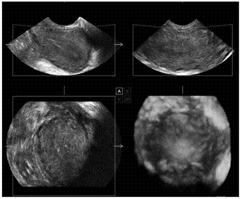
[0031] S101. Acquire a 3D ultrasound image of the uterus including endometrium.

[0032] The operator holds the probe in hand, transmits ultrasonic waves to the uterus of the person to be checked through the probe, and receives the ultrasonic beams transmitted back from the uterus, and sends them to the image processor after beam synthesis to form an ultrasonic image.

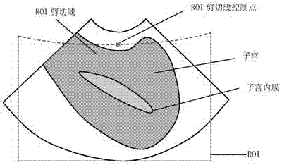
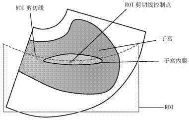
[0033] Such as Figure 7 As shown, the 3D ultrasound image includes: A section, B section, C section, and 3D image. The slices A, B, and C are slice images along the X-axis, Y-axis, and Z-axis, respectively, where the planes A and B are often used as planes for endometrial observation.

[0034] The ultrasonic image of the uterus includes, from the center to the outside, a cavity at the center, an endometrium surrounding the cavity, an envelope outside the endometrium, tissues outside the envelope, and a uterine wall at the outermost layer of the tissue. When judging whether there is an abnormality in the uter...

Embodiment 2

[0054] In some other embodiments, in order to enhance the accuracy of endometrial image extraction, the step of image enhancement on the 3D ultrasound image of the uterus is also included before the extraction of the endometrial image, so as to better help extract the uterine Intima image.

[0055] In order to emphasize the two boundary lines of the endometrium, make the original blurry boundary image clear and continuous, and suppress uninteresting features, we use image enhancement to process the endometrium, the main purpose is to remove noise and enhance edge.

[0056] The A plane in the 3D view of the uterus was selected as the reference plane, and the two-dimensional anisotropic filter was used to denoise the image to make the image more uniform. Then adjust the image contrast to make the endometrium easier to observe: statistical image gray value (removing zero data) to obtain a histogram, determine the two inflection points of the image for gray stretching; use piecewis...

Embodiment 3

[0067] In some embodiments, in order to increase the accuracy of extracting the endometrial image, before performing step S102 to extract the endometrial image, the approximate range of the endometrial image may be determined according to the received signal input by the operator. For example, in the area where the endometrium can be determined by the naked eye, the endometrium part is roughly marked by drawing points, lines, and frames, thereby improving the accuracy of automatically extracting the endometrium image. That is, through the semi-automatic method, the accuracy of endometrial image extraction is increased, and the accuracy of subsequent operations is also improved. The method as a whole may then include the following:

[0068] S301. Acquire a 3D ultrasound image of the uterus including endometrium.

[0069] S302. Perform image enhancement on the 3D ultrasound image of the uterus.

[0070] S303. Determine the approximate range of the endometrial image according t...


PUM


Abstract

The invention provides an ultrasonic imaging method used for carrying out 3D imaging on the endometrium. The method includes the steps that a uterus 3D ultrasonic image containing the endometrium is acquired; the ultrasonic image is rotated so that the endometrium part in the ultrasonic image can be rotated to be horizontal; an ROI shear line is adjusted to axially correspond to the endometrium image; an endometrium 3D image extracted according to the adjusted ROI shear line is acquired. The invention further provides a corresponding device and ultrasonic equipment. By the adoption of the method, after a doctor acquires the uterus 3D ultrasonic image containing the endometrium, the ultrasonic image is rotated to make the endometrium image horizontal, then the shear line is adjusted to axially correspond to the endometrium, and thus the endometrium 3D image is automatically acquired; operation time is greatly shortened, and the operation process is simplified.

Description

technical field [0001] The invention relates to the technical field of ultrasonic imaging, in particular to an ultrasonic imaging method, device and ultrasonic equipment. Background technique [0002] The traditional three-dimensional image observation of the endometrium is obtained by the doctor by manually adjusting the image. Due to the large individual differences, especially the endometrium of postpartum women has not fully recovered, the line echo of the endometrium is relatively close to the echo of other tissues, and the doctor is very careful. Difficult to distinguish and adjust a better cut line to fit the morphology of the endometrial borderline. And it is relatively time-consuming to rotate the image and adjust the cutting line. Contents of the invention [0003] The present invention provides an ultrasonic imaging method for performing 3D imaging on the endometrium, the method comprising: [0004] Obtain a 3D ultrasound image of the uterus including the endo...

Claims


Application Information

Patent Timeline
IPC IPC(8): A61B8/00
Inventor 唐艳红艾金钦刘旭江
Owner SONOSCAPE MEDICAL CORP
Why Eureka
Try Eureka