Method of fully automatically classifying and partitioning branch retinal artery obstruction based on three-dimensional OCT image

An arterial occlusion and retinal technology, applied in image analysis, image enhancement, image data processing, etc., can solve the problems of no analysis, inability to provide quantitative information on the occlusion area, and no classification of retinal branch artery occlusion, etc., to achieve high accuracy. Effect

Active Publication Date: 2016-05-04
SUZHOU BIGVISION MEDICAL TECH CO LTD
View PDF3 Cites 13 Cited by
  • Summary
  • Abstract
  • Description
  • Claims
  • Application Information

AI Technical Summary

Problems solved by technology

[0004] These methods are all qualitative analysis of branch retinal artery occlusion and cannot detect and segment the occluded area fully automatically
Therefore, it cannot provide clinicians with accurate quantitative information about the obstructed area, such as shape, size and location, etc.
In general, the current methods for branch retinal artery occlusion have the following defects: (1) most algorithms do not classify branch retinal artery occlusion (acute phase and atrophic phase), and the organizational structure of retina in different stages of branch retinal artery occlusion A big difference
(2) Most methods are not fully automatic, relying on manual measurement or marking
(3) Most of the algorithms do not perform a specific analysis of the occlusion area of ​​branch retinal artery occlusion
However, the shape, size, and location of the blocked area of ​​retinal branch artery occlusion are arbitrary, and the boundary between the blocked area and surrounding tissues is very blurred, and the retinal OCT image itself is noisy.
Therefore, fully automated segmentation of branch retinal artery occluded regions is a challenging task

Method used


Image

  • Method of fully automatically classifying and partitioning branch retinal artery obstruction based on three-dimensional OCT image
  • Method of fully automatically classifying and partitioning branch retinal artery obstruction based on three-dimensional OCT image
  • Method of fully automatically classifying and partitioning branch retinal artery obstruction based on three-dimensional OCT image

Examples


Embodiment Construction

[0046] In order to make the technical means, creative features, goals and effects achieved by the present invention easy to understand, the present invention will be further described below in conjunction with specific embodiments.

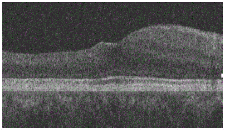
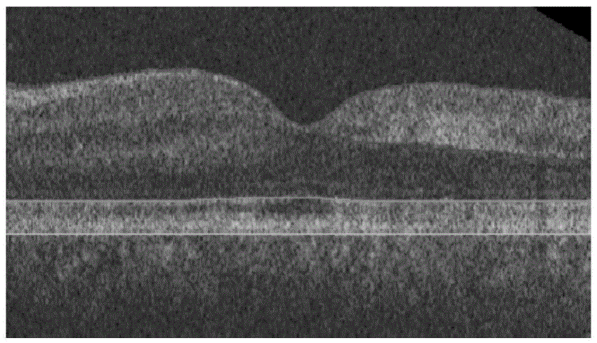
[0047] The invention comprises the following four steps: image preprocessing, classification based on AdaBosst, segmentation of the acute stage of branch retinal artery occlusion and segmentation of the atrophy stage of branch retinal artery occlusion.

[0048] (1) Image preprocessing

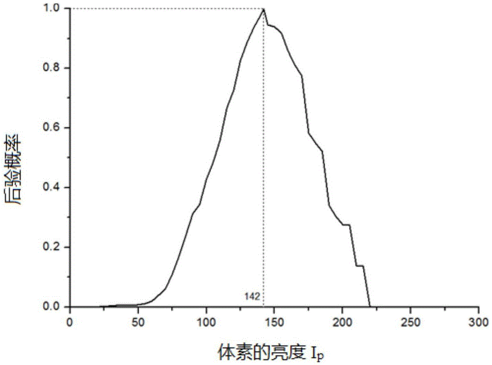
[0049] In order to obtain the information of each layer of the retina, a graph search method is used to realize the layering of the retina. Its cost function is defined as:

[0050] E ( S ) = Σ v ∈ S c v + Σ ( p ...


PUM


Abstract

The invention discloses a method of fully automatically classifying and partitioning a branch retinal artery obstruction based on a three-dimensional OCT image. The method comprises the following steps: pretreatment is carried out, a graph searching algorithm is adopted to layer the retina, and each layer of the retina is leveled according to a pigment epithelium layer; an AdaBoost classifier is used for automatically classifying an acute stage and an atrophy stage of the branch retinal artery obstruction; partitioning of the acute stage of the branch retinal artery obstruction is carried out, a Bayesian posterior probability is firstly adopted to carry out initial partitioning on an obstruction area, and then based on a graph searching-graph partitioning algorithm, accurate partitioning is carried out on the obstruction area; and partitioning of the atrophy stage of the branch retinal artery obstruction is carried out, and the obstruction area for the atrophy stage is automatically partitioned through building an inner retina thickness model. The method of the invention can accurately classify and partition the branch retinal artery obstruction area, and can replace manual classifying and partitioning.

Description

technical field [0001] The present invention relates to the classification of lesions in retinal images of SD-OCT (frequency-domain optical coherence tomography) and the segmentation method of lesion areas, in particular to a method for fully automatic classification and segmentation of retinal branch artery occlusion based on three-dimensional OCT images, belonging to Method for classifying and segmenting retinal images Technical field. Background technique [0002] Branch retinal artery occlusion is one of the acute diseases in ophthalmology. It has a poor prognosis, rapid onset, and usually painless monocular visual impairment. Retinal artery occlusion interrupts the nutrient supply of the corresponding retinal area, leading to hypoxia and ischemia in the local area of ​​the retina, resulting in edema and rapid death of retinal cells, resulting in visual dysfunction. [0003] So far, most of the work related to branch retinal artery occlusion has focused on the qualitat...

Claims


Application Information

Patent Timeline
Patent Type & Authority Applications(China)
IPC IPC(8): G06T7/00
CPCG06T2207/10101G06T2207/20081G06T2207/30041
Inventor 陈新建郭静云朱伟芳陈浩宇
Owner SUZHOU BIGVISION MEDICAL TECH CO LTD
Why Eureka
Try Eureka