Extraction method of skin area in medical image

A skin area and medical image technology, applied in the field of medical image processing, can solve problems such as complex processing, time-consuming, and not well applicable to clinical diagnosis, and achieve fast and simple effects

Inactive Publication Date: 2016-05-18
ZHUHAI HOKAI MEDICAL INSTR +1
View PDF4 Cites 4 Cited by
  • Summary
  • Abstract
  • Description
  • Claims
  • Application Information

AI Technical Summary

Problems solved by technology

Although this method can automatically extract the patient's skin area, the processing is complicated and time-consuming, and it is not well suitable for actual clinical diagnosis.

Method used


Image

  • Extraction method of skin area in medical image
  • Extraction method of skin area in medical image
  • Extraction method of skin area in medical image

Examples


Embodiment Construction

[0026] In order to make the above and other objects, features and advantages of the present invention more apparent, the following specifically cites the embodiments of the present invention, together with the accompanying drawings, for a detailed description as follows.

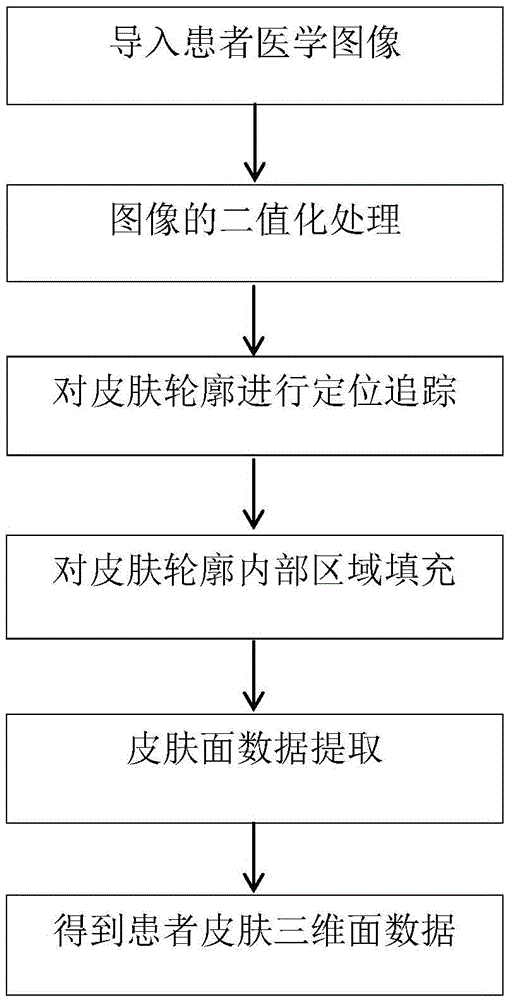
[0027] refer to figure 1 , figure 1 It is a flow chart of the method of the present invention, and the steps of the method for extracting the skin region in the medical image of the present invention are as follows:

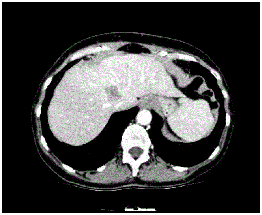
[0028] Step 1. Import the medical image of the patient to obtain a sequence slice image. The width of the sequence slice image is width and the height is height. The sequence slice image is as follows figure 2 shown;

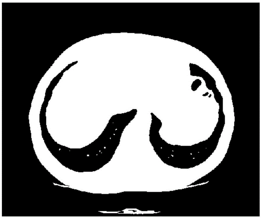
[0029] Step 2, carry out binarization processing on sequence slice image, obtain sequence binarization slice image; Adopt OTSU algorithm to carry out binarization processing to image in this embodiment, the image after binarization processing is as follows image 3 shown;

...


PUM


Abstract

The invention provides an extraction method of a skin area in a medical image. The extraction method comprises the following steps of: importing the medical image of a patient, obtaining a sequence slice image; carrying out binary processing on the sequence slice image; carrying out high gray value point searching on the sequence binary slice image, and when a high gray value point is searched, judging whether the high gray value point is and isolated point, if yes, continuing searching, if not, carrying out contour tracking with the high gray value point serving as a start point, obtaining the number of contour point sets, then judging whether the number of contour point sets is larger than a preset threshold, if yes, indicating that skin area contour extraction is successful, if not, continuing searching for the next high gray value point until the number of obtained contour point sets is larger than the preset threshold, and obtaining a sequence skin contour image; carrying out area filling on the sequence skin contour image; and carrying out face extraction on the sequence skin contour image, obtaining three-dimensional face data of the skin area, and completing the extraction of the skin area. The method is simple to realize, high in speed and capable of meeting a plurality of clinic demands.

Description

technical field [0001] The invention relates to a medical image processing method, in particular to a method for extracting a skin region in a medical image. Background technique [0002] The extraction of the patient's skin area plays an important role in percutaneous puncture surgery. The accuracy of skin area extraction directly affects the selection of the needle insertion point, the planning of the needle insertion path, and then affects the entire subsequent puncture process. In the current medical image processing software, the extraction of patients' skin areas generally requires manual intervention by users, such as setting thresholds, deleting non-skin areas, etc., and the extraction methods are time-consuming and cumbersome. The Chinese invention patent application with publication number 1555028 discloses an automatic skin extraction method in medical image segmentation. This method needs to establish different mathematical models of skin tissue according to the ...

Claims


Application Information

Patent Timeline
IPC IPC(8): G06T7/00
CPCG06T7/0012G06T2207/30088
Inventor 陈庆辉叶鹏飞谢波陈钢梁萍程志刚
Owner ZHUHAI HOKAI MEDICAL INSTR
Why Eureka
Try Eureka