Body position icon, adding method, control device and ultrasonic equipment of control device
A body position map, ultrasound technology, applied in ultrasonic/sonic/infrasonic diagnosis, sonic diagnosis, infrasonic diagnosis and other directions, can solve problems such as unfavorable
- Summary
- Abstract
- Description
- Claims
- Application Information
AI Technical Summary
Problems solved by technology
Method used
Image
Examples
Embodiment 1
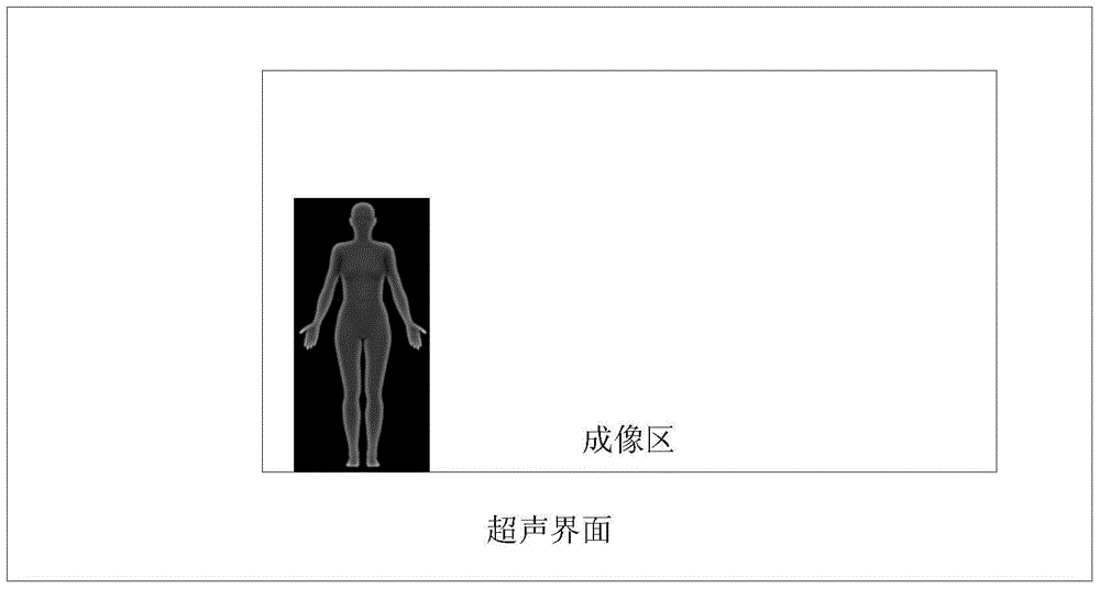
[0029] When performing ultrasound examination on various parts of the human body, it is usually necessary to add a body position map mark on the ultrasound image display interface to mark the examination site, so as to facilitate the doctor's identification of the examination site. The existing body position icons are flat, but the human body is usually three-dimensional, so it is not conducive to accurately reflect the specific orientation of the inspection.
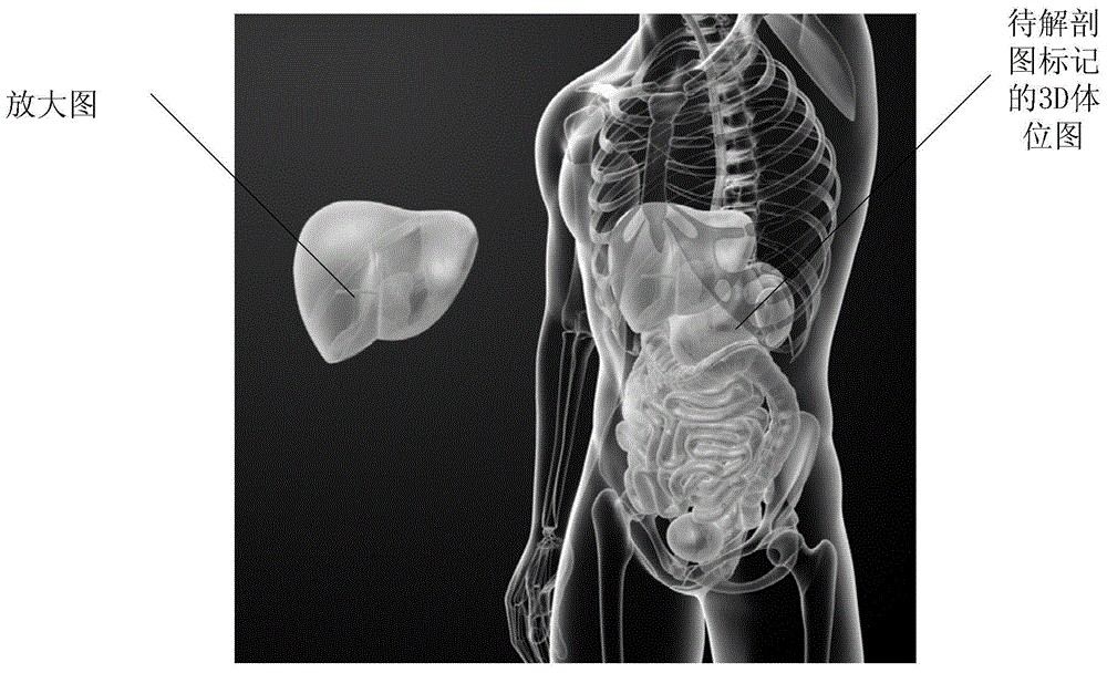
[0030] Such as figure 1 As shown, the present invention provides a body position map, and the body position map is a 3D body position map.
[0031] The body position map may be displayed on the ultrasound imaging interface by default, or a 3D body position map of a human body may be added to the ultrasound imaging interface by clicking the [body marker] button. Usually, the body position diagram also includes a probe mark, and different positions of the human body where the probe is located can be identified by the pos...
Embodiment 2
[0045] The present invention also provides a method for adding a body position map described in Embodiment 1, the method includes the following steps:
[0046] S101. Rotate the body position map until it is consistent with the current body position of the examinee.
[0047] The body position map may be displayed on the ultrasound imaging interface by default, or a 3D body position map of a human body may appear on the ultrasound imaging interface by clicking the [body mark] button, and various marks may also be included on the 3D body position map, For details, refer to the relevant description of the first embodiment.
[0048] At this time, the probe mark on the body position chart rotates synchronously with the body position chart.
[0049] Control the rotation of the body position map through the control unit (such as a trackball, direction keys), until it is consistent with the position of the currently examined patient (such as lying on the stomach, sideways, lying down,...
Embodiment 3
[0060] As shown in the figure, the present invention also provides a control device 200 for a body position map or a method for adding a body position map as described in Embodiment 1 or 2, wherein the device 200 includes: a display unit 201, a storage unit 202 , the first control unit 203;
[0061] The storage unit 202 is configured to store the volume 3D bitmap;
[0062] The first control unit 203 is configured to send the 3D body position map stored in the storage unit to the display unit 201 for display.
[0063] In some embodiments, the device further includes: a first input unit 204, a second control unit 205;
[0064] The first input unit is used for inputting a control signal for rotating the body position map; the input unit may include: a trackball, an arrow key, and the like. For example, if the trackball rotates clockwise or counterclockwise, it can represent the clockwise or counterclockwise rotation of the body position map. Specifically, you can set how many d...
PUM
 Abstract
Description
Claims
Application Information
 - R&D
- Intellectual Property
- Life Sciences
- Materials
- Tech Scout
- Unparalleled Data Quality
- Higher Quality Content
- 60% Fewer Hallucinations
Browse by: Latest US Patents, China's latest patents, Technical Efficacy Thesaurus, Application Domain, Technology Topic, Popular Technical Reports.
© 2025 PatSnap. All rights reserved.Legal|Privacy policy|Modern Slavery Act Transparency Statement|Sitemap|About US| Contact US: help@patsnap.com


