Automatic medical image interlayer sketching method, device and system

A technology for medical images and images, applied in the field of medical instruments, can solve the problems of large amount of registration calculation, poor registration accuracy, and time-consuming automatic delineation, so as to reduce noise interference, reduce the risk of human error, and improve automatic delineation. effect of effect

Inactive Publication Date: 2016-07-20
SHENZHEN YINO INTELLIGENCE TECH
View PDF3 Cites 6 Cited by
  • Summary
  • Abstract
  • Description
  • Claims
  • Application Information

AI Technical Summary

Problems solved by technology
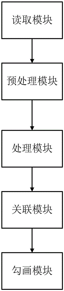
The disadvantage of this type of method is that a suitable prior data set needs to be prepared in the early stage. On the other hand, there is a poor registration accuracy caused by the uncertainty of the optimal template, and the large amount of registration calculations leads to a long time-consuming automatic delineation.

Method used


Image

  • Automatic medical image interlayer sketching method, device and system
  • Automatic medical image interlayer sketching method, device and system
  • Automatic medical image interlayer sketching method, device and system

Examples


no. 1 example

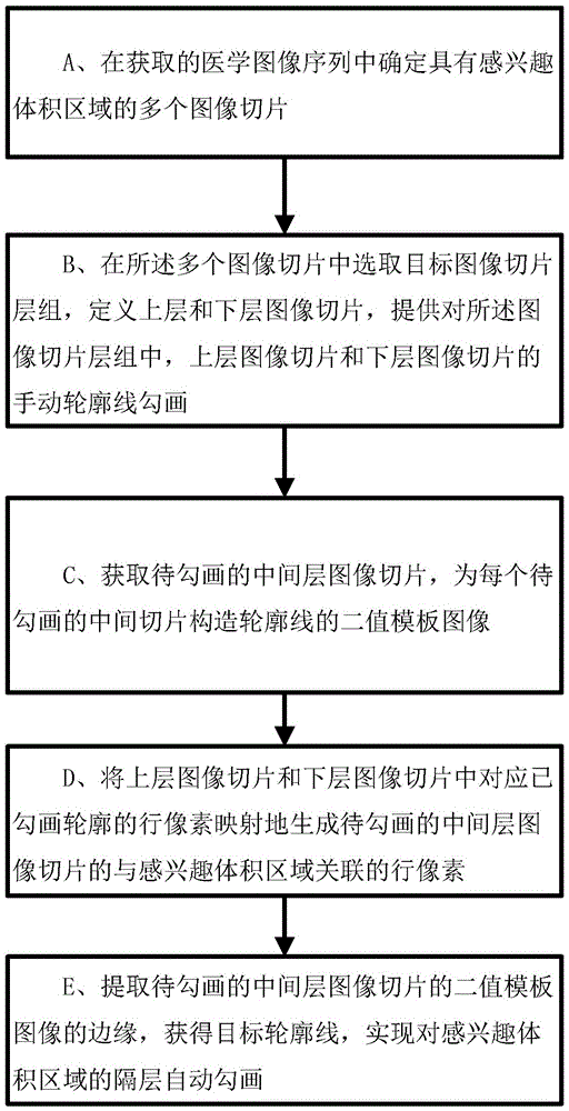
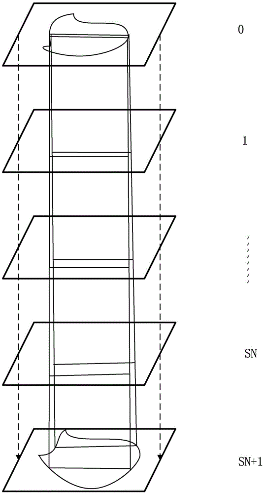
[0025] Based on the first embodiment of the invention, such as figure 1 A method for automatically delineating medical image compartments is shown, comprising the following steps:

[0026] A. Determining a plurality of image slices with volume regions of interest in the acquired medical image sequence; B. Selecting a target image slice layer group among the plurality of image slices, defining upper and lower layer image slices, and providing a description of the image In the slice layer group, the manual outline drawing of the upper layer image slice and the lower layer image slice; C. Obtain all the middle layer image slices to be drawn, and construct a binary template image of the outline for each middle slice to be drawn; D. The line pixels corresponding to the outline in the upper layer image slice and the lower layer image slice use the image pixel space coordinate projection method to map to generate the row pixels associated with the volume of interest region of the mid...


PUM


Abstract

The invention discloses an automatic medical image interlayer sketching method, device and system. The method comprises the steps of: obtaining a plurality of image sections having interested volume areas; selecting a target image section layer set, defining upper and lower image sections and providing manual outline sketching; obtaining an interlayer image section, and constructing a two-value template image of the outline; generating row pixels associated with the interested volume areas in a mapping manner by using the row pixels corresponding to sketched outline in the upper and lower image sections; and extracting edges of the two-value template image to obtain the target outline. The device comprises a reading module, a preprocessing module, a processing module, a correlation module and a sketching module. The system comprises corresponding computer hardware to execute the above method. The automatic medical image interlayer sketching method, device and system have the advantages that the noise disturbance is lowered, the automatic sketching effect of areas with undefined edges is improved, the dependence on the prophase sketching and the target area data is reduced, the rapid realization of the automatic sketching is facilitated, and the risk of human errors is lowered.

Description

technical field [0001] The invention relates to a method, device and system for automatically delineating medical image compartments, belonging to the field of medical instruments. Background technique [0002] In the field of tumor radiotherapy, after the radiologist arranges a radiotherapy plan based on radiation exposure for tumor patients, the radiotherapy physicist designs a radiotherapy plan that meets the requirements of the treatment plan. Among them, the volume of interest (VOI), which is used to describe the patient's organs at risk and the target target area, is outlined by the radiotherapist in CT and MR images, which is a key step in the radiotherapy plan, which directly determines the accuracy of the radiotherapy plan. . [0003] Usually, volume regions of interest such as organs at risk and target volumes are composed of a set of contour lines, which are distributed on multiple image slices. In order to ensure precision and accuracy, radiotherapy doctors or ...

Claims


Application Information

Patent Timeline
Patent Type & Authority Applications(China)
IPC IPC(8): G06T11/00
CPCG06T11/006G06T2211/432
Inventor 刘常春吴江华李培强
Owner SHENZHEN YINO INTELLIGENCE TECH
Why Eureka
Try Eureka