Cup type time-resolved fluorescence D-dimer analysis method and reagent kit based on microspheres

A technology of time-resolved fluorescence and analysis method, which is applied in the field of cup type time-resolved fluorescence D-dimer analysis method and kit based on microspheres, which can solve the problem of unsatisfactory D-dimer detection precision and complicated detection steps , poor precision and other problems, to achieve the effect of reducing steric hindrance effect, shortening detection time and improving detection sensitivity

Inactive Publication Date: 2016-08-24
SHANGHAI UPPER BIO TECH PHARMA
View PDF6 Cites 4 Cited by
  • Summary
  • Abstract
  • Description
  • Claims
  • Application Information

AI Technical Summary

Problems solved by technology
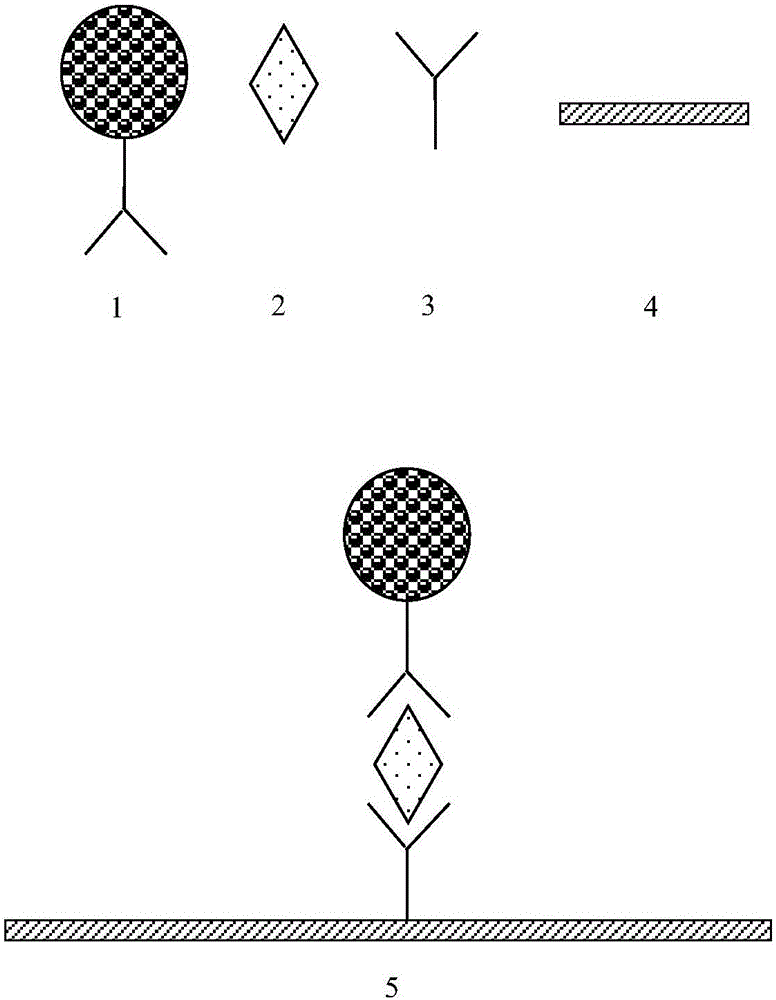
[0007] The purpose of the present invention is to overcome the shortcomings of the above-mentioned existing fluorescent immunochromatography method for measuring D-dimer detection precision is not ideal and the time-resolved fluorescence detection method needs a dissociation enhancement process, the detection steps are complicated, the time is long, and the accuracy is poor. A microsphere-based cup-type time-resolved fluorescent D-dimer analysis method and kit

Method used


Image

  • Cup type time-resolved fluorescence D-dimer analysis method and reagent kit based on microspheres
  • Cup type time-resolved fluorescence D-dimer analysis method and reagent kit based on microspheres
  • Cup type time-resolved fluorescence D-dimer analysis method and reagent kit based on microspheres

Examples


Embodiment 1

[0055] 1. Preparation of detection cuvette

[0056] (1) Detection cuvette coating

[0057] D-dimer antibody 1401 (Medix Company) was diluted to 40 μg / ml with 0.2M phosphate buffer (pH 7.8), 100 μL / well, coated at 37° C. for 4 hours, and washed.

[0058] (2) Detect cuvette closure

[0059] Add 200 μL / well of blocking buffer to the reaction cup, block at 37°C for 4 hours, discard the blocking solution in the coated reaction cup, and pat dry.

[0060] (3) Detection of cuvette drying

[0061] The sealed cuvette was placed in a 37° C. drying oven with a humidity lower than 30% for 8 hours, sealed and dried for storage.

[0062] 2. Preparation of fluorescently labeled antibodies

[0063] (1) Pretreatment of fluorescent particles

[0064] Take 1mg of carboxyl time-resolved fluorescent microspheres (200nm, 0.1ml, 10mg / mL, Bangslab company), add 60ul 500mM MES (2-(N-morpholine)ethanesulfonic acid) buffer, pH 6.0, add 0.2mg 1 -(3-Dimethylaminopropyl)-3-ethylcarbodiimide hydrochlor...

Embodiment 2

[0091] (1) Detection cuvette coating

[0092] D-dimer antibody 1401 (Medix Company) was diluted to 40 μg / ml with 0.2M phosphate buffer (pH 7.8), 100 μL / well, coated at 37° C. for 4 hours, and washed.

[0093] (2) Detect cuvette closure

[0094] Add 200 μL / well of blocking buffer to the reaction cup, block at 37°C for 4 hours, discard the blocking solution in the coated reaction cup, and pat dry.

[0095] (3) Detection of cuvette drying

[0096] The sealed cuvette was placed in a 37° C. drying oven with a humidity lower than 30% for 8 hours, sealed and dried for storage.

[0097] 2. Preparation of fluorescently labeled antibodies

[0098] (1) Pretreatment of fluorescent particles

[0099] Take 1mg of carboxyl time-resolved fluorescent microspheres (200nm, 0.1ml, 10mg / mL, Bangslab company), add 60ul 500mM MES (2-(N-morpholine)ethanesulfonic acid) buffer, pH 6.0, add 0.2mg 1 -(3-Dimethylaminopropyl)-3-ethylcarbodiimide hydrochloride (EDC), add 0.4mg N-hydroxysuccinimide (NHS...


PUM

PropertyMeasurementUnit
particle sizeaaaaaaaaaa
particle diameteraaaaaaaaaa

Abstract

The invention discloses a cup type time-resolved fluorescence D-dimer analysis method and a reagent kit based on microspheres. The method includes the steps that the inner surface of a detection reaction cup is coated with D-dimer monoclonal antibodies or polyclonal antibodies; the time-resolved fluorescence microspheres and the D-dimer monoclonal antibodies or polyclonal antibodies are subjected to covalent bond cross-linking to prepare fluorescence-labeled antibodies; a D-dimer sample to be detected is added into the coated detection reaction cup, then the fluorescence-labeled antibodies are added, and incubation is carried out at 25-40 DEG C; cleaning is carried out to remove unbound antigens and fluorescence-labeled antibodies, and fluorescence signals in the detection reaction cup are tested under the excitation of 340-380 nm exciting light; the fluorescence signals are compared with a standard curve, and the D-dimer concentration of the D-dimer sample to be detected is obtained. By means of the method, detection can be completed within a short detection time, the detection time is shortened, and the detection sensitivity is effectively improved.

Description

technical field [0001] The invention relates to the technical field of D-dimer detection, in particular to a microsphere-based cup-type time-resolved fluorescence D-dimer analysis method and a kit. Background technique [0002] D-dimer (D-dimer, D-D) is a specific degradation product of cross-linked fibrin (Fb). The fibrinolysis system is the most important anticoagulant system in the human body. When a fibrin clot is formed, in the presence of plasminogen activator, plasminogen is activated and converted into plasmin , the process of fibrinolysis begins, plasmin degrades fibrin clots to form various soluble fragments, forming fibrin product (FDP), FDP is composed of the following substances: X-oligomer (X-oligomer), D-dimer (D-Dimer), intermediate fragments (Intermediate fragments), fragment E (Fragment E). Among the degradation products of fibrinolytic protein, only D-dimer cross-linked fragments can reflect the thrombolytic activity after thrombus formation. Therefore,...

Claims


Application Information

Patent Timeline
Patent Type & Authority Applications(China)
IPC IPC(8): G01N21/64G01N33/58
CPCG01N21/6408G01N33/582G01N33/585
Inventor 石晓强杨晶周奕璇李福刚徐建新
Owner SHANGHAI UPPER BIO TECH PHARMA
Why Eureka
Try Eureka