Microballoon-based cup-type time resolution fluorescent procalcitonin analysis kit, preparation method and application thereof

A technology of time-resolved fluorescence and fluorescent calcitonin, which is applied in the field of clinical medical diagnosis, can solve the problems of high technical requirements of electrochemiluminescence, high technical requirements of chemiluminescence, and unsatisfactory detection precision, and achieve easy automatic operation, The effect of shortening the detection time and reducing the effect of steric hindrance

Inactive Publication Date: 2016-07-13
SHANGHAI UPPER BIO TECH PHARMA
View PDF7 Cites 4 Cited by
  • Summary
  • Abstract
  • Description
  • Claims
  • Application Information

AI Technical Summary

Problems solved by technology
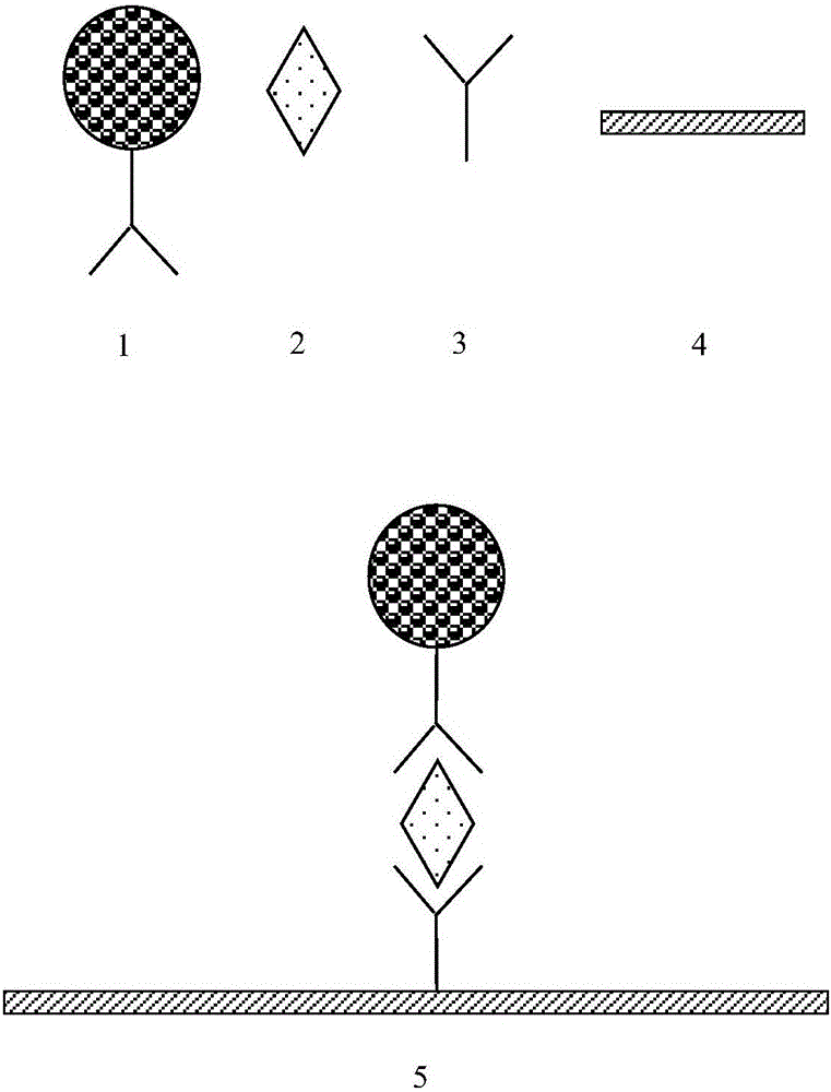
However, radioimmunoassay and enzyme-linked immunosorbent assay are complicated to operate and take a long time to detect, so they have almost been eliminated at present; chemiluminescence method has high technical requirements, and the operation steps are relatively cumbersome, requiring multi-step reagent addition process; High requirements, not easy to carry out routinely in clinical laboratories
Although the colloidal gold immunochromatography method has the advantages of less sample consumption, simple, quick, and cheap, however, when the antigen or antibody content in some samples is extremely low, the color of the colloidal gold will be very light or even no color, and it is difficult to Judging the results with the naked eye is prone to misjudgment and low sensitivity
Although the sensitivity of fluorescence immunochromatography is higher than that of colloidal gold immunochromatography, its detection precision is not ideal, and the sensitivity is still far behind that of large-scale chemiluminescence or electrochemiluminescence instruments

Method used


Image

  • Microballoon-based cup-type time resolution fluorescent procalcitonin analysis kit, preparation method and application thereof
  • Microballoon-based cup-type time resolution fluorescent procalcitonin analysis kit, preparation method and application thereof
  • Microballoon-based cup-type time resolution fluorescent procalcitonin analysis kit, preparation method and application thereof

Examples


Embodiment 1

[0048] (1) Preparation of detection cuvette:

[0049] A. Detection cuvette coating: Procalcitonin antibody 18B7 (Hytest Company) was diluted to 10 μg / ml with 0.2mol / L phosphate buffer (pH7.8), 100 μL / well, coated at 37°C for 4 hours, washed plate.

[0050] B. Detection cuvette blocking: Add 200 μL / well of blocking buffer to the cuvette, block at 37°C for 4 hours, discard the blocking solution in the coated cuvette, and pat dry.

[0051] C. Drying of the test cuvette: place the sealed cuvette above in a 37° C. drying oven with a humidity lower than 30% for 4 hours, and store in a sealed and dry place.

[0052] (2) Preparation of fluorescently labeled antibodies:

[0053] A. Fluorescent particle activation:

[0054] Take 1mg of carboxyl time-resolved fluorescent microspheres (200nm, 0.1ml, 10mg / mL, Bangslab Company), add 60μL of 500mmol / L MES (2-(N-morpholine)ethanesulfonic acid) buffer, pH6.0, add 0.2 mg1-(3-dimethylaminopropyl)-3-ethylcarbodiimide hydrochloride (EDC), add ...

Embodiment 2

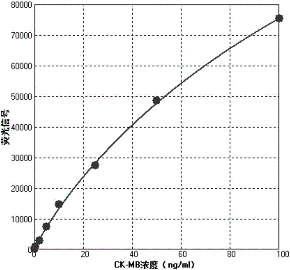
[0064] (1) Drawing of standard curve:

[0065] The reagents prepared in Example 1 were used to form a procalcitonin time-resolved fluorescence quantitative kit, and the calibrator was measured, and each concentration was repeated 10 times.

[0066] Add 10 μL of calibrator and 50 μL of fluorescently labeled antibody to each test, incubate at 37°C for 20 min, then wash the detection cup, and read on the VictorX4 fluorescence reader from PerkinElmer (excitation wavelength 340-380nm, detection wavelength 600-630nm). The data are shown in Table 1.

[0067] Table 1

[0068]

[0069] According to the data in Table 1, a standard curve was drawn with the concentration of the calibrator as the abscissa and the mean fluorescence signal as the ordinate. standard curve as figure 2 shown. The standard curve has good linearity, and the concentration of procalcitonin contained in the sample can be quantitatively analyzed by the standard curve.

[0070] It can be seen from the results...

Embodiment 3

[0078] (1) Preparation of detection cuvette:

[0079] A. Detection cuvette coating: Procalcitonin antibody 18B7 (Hytest Company) was diluted to 10 μg / ml with 0.2mol / L phosphate buffer (pH7.8), 100 μL / well, coated at 37°C for 4 hours, washed plate.

[0080] B. Detection cuvette blocking: Add 200 μL / well of blocking buffer to the cuvette, block at 37°C for 4 hours, discard the blocking solution in the coated cuvette, and pat dry.

[0081] C. Detection of drying of the cuvette: place the sealed cuvette in a drying oven at 37° C. with a humidity lower than 30% for 4 hours, and store in a sealed and dry place.

[0082] (2) Preparation of fluorescently labeled antibodies:

[0083] A. Fluorescent particle activation:

[0084] Take 1mg of carboxyl time-resolved fluorescent microspheres (200nm, 0.1ml, 10mg / mL, Bangslab company), add 60ul 500mMMES (2-(N-morpholine)ethanesulfonic acid) buffer, pH 6.0, add 0.2mg1-(3 -Dimethylaminopropyl)-3-ethylcarbodiimide hydrochloride (EDC), add 0....


PUM

PropertyMeasurementUnit
particle diameteraaaaaaaaaa

Abstract

The invention belongs to the field of clinical medical diagnosis and especially relates to a microballoon-based cup-type time resolution fluorescent procalcitonin analysis kit, a preparation method and an application thereof. The kit comprises a detection reaction cup, a fluorescence labeling antibody and a cleaning fluid. The method comprises the following steps of: in specific detection process, firstly drawing a standard curve; adding a to-be-detected sample into the detection reaction cup; adding the fluorescence labeling antibody; incubating at 30-40 DEG C; washing with the cleaning fluid and removing the uncombined antibody and fluorescence labeling antibody; and comparing a fluorescence signal with the standard curve, thereby acquiring the concentration of procalcitonin of the to-be-detected sample. The detection method for the procalcitonin is ultrahigh in sensitivity, so that the automatic operation can be easily realized; the reaction temperature is uniform; the method is not influenced by the environmental factor; the accuracy is better; the precision is excellent.

Description

technical field [0001] The invention belongs to the field of clinical medical diagnosis, and in particular relates to a cup-type time-resolved fluorescent procalcitonin analysis kit based on microspheres and a preparation method and application thereof. Background technique [0002] Procalcitonin (PCT) is a non-invasive clinical laboratory index used for the diagnosis and treatment monitoring of severe bacterial infections. Similar studies have shown that PCT is increasingly recognized as a good marker of bacterial infection and sepsis, thus becoming an important tool in clinical diagnosis. Increases in PCT concentrations, reflecting the progression from a healthy state to the most severe consequences of bacterial infection (sepsis, severe sepsis, septic shock), showed a positive correlation. Therefore, for bacterial infection and sepsis, high-sensitivity and full-quantitative PCT detection can not only perform early clinical diagnosis, but also indicate the disease process...

Claims


Application Information

Patent Timeline
Patent Type & Authority Applications(China)
IPC IPC(8): G01N21/64G01N33/577G01N33/533
CPCG01N21/6486G01N33/533G01N33/577
Inventor 石晓强李福刚徐建新周奕璇杨晶
Owner SHANGHAI UPPER BIO TECH PHARMA
Why Eureka
Try Eureka