Two-dimensional image and CT or MR image three-dimensional fusion method

A two-dimensional image, three-dimensional fusion technology, applied in the field of digital medical imaging engineering, can solve problems such as deficiencies

Active Publication Date: 2016-12-07
THE FIRST AFFILIATED HOSPITAL OF XIAN JIAOTONG UNIV
View PDF5 Cites 21 Cited by
  • Summary
  • Abstract
  • Description
  • Claims
  • Application Information

AI Technical Summary

Problems solved by technology

Although this method preliminarily realizes the infrared thermal Figure three 3D positioning, but registration and fusion methods based on marker points

Method used


Image

  • Two-dimensional image and CT or MR image three-dimensional fusion method
  • Two-dimensional image and CT or MR image three-dimensional fusion method
  • Two-dimensional image and CT or MR image three-dimensional fusion method

Examples


Embodiment 1

[0061] In the following embodiments, the technical solutions of the present invention will be described in detail by taking the three-dimensional fusion of two-dimensional infrared thermal images and MR images from three perspectives (left side, front side and right side) as an example.

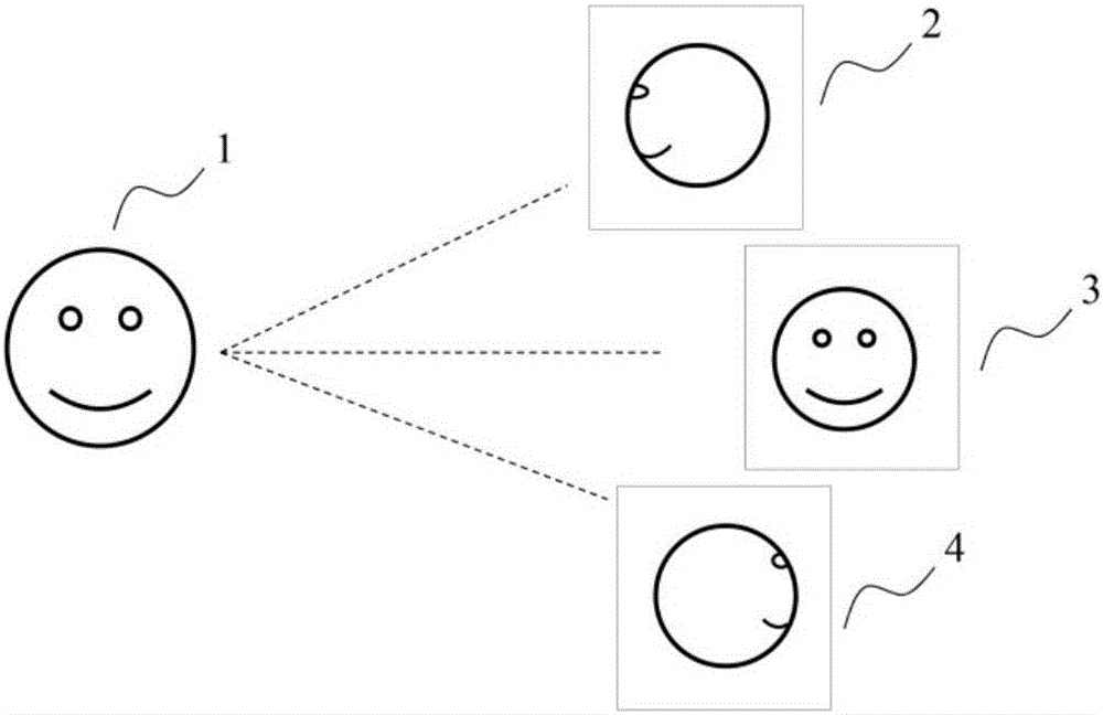
[0062] (1) if figure 1 As shown, for the reconstructed part 1, the three-view two-dimensional infrared thermal image acquisition is carried out, and three infrared thermal images of the left side 2, the front side 3 and the right side 4 are respectively obtained.

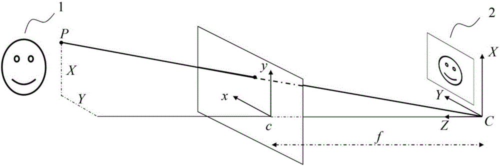
[0063] (2) if figure 2 As shown, the 3D geometric structure 1 is reconstructed based on the MR image of the reconstructed part; the 3D geometric structure 1 is virtually imaged according to the principle of pinhole imaging, and the 2D plane projection image 2 with the same acquisition angle as the 2D infrared heat map is obtained through calculation.

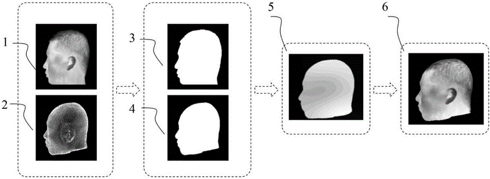
[0064] (4) if image 3 As shown, the image of the left face of the human fac...

Embodiment 2

[0068] Different from Embodiment 1, in this embodiment, two-dimensional images of more viewing angles can also be collected, for example, within a range of 360° centering on the object to be reconstructed, one target image is collected for each 30°, a total of 12 images. Two-dimensional projection images from 12 viewing angles should also be obtained corresponding to the three-dimensional CT or MR geometry.

Embodiment 3

[0070] Different from Embodiment 1, the MR images in this embodiment can be completely replaced by CT images.


PUM


Abstract

The invention discloses a two-dimensional image and CR or MR image three-dimensional fusion method. The method comprises the steps that three-dimensional geometric structure information of a CT or MRI structure image is used for combining multi-angle two-dimensional images for human body three-dimensional target image reconstruction, and then target image and MR image three-dimensional multi-modal fusion is achieved. According to the three-dimensional medical image fusion technology, the purpose of two-dimensional image three-dimensional complex structure positioning is achieved, and more precise focus positioning is provided; in addition, the two-dimensional image and the CT or MR image are subjected to three-dimensional fusion, multi-modal image system construction is achieved, more abundant resource information is provided for clinical disease diagnosis, and the diagnostic efficiency is improved.

Description

Technical field: [0001] The invention relates to a three-dimensional fusion method of a two-dimensional image and a traditional medical image of computerized tomography (CT) and magnetic resonance image (Magnetic Resonance Imaging, MRI), belonging to the technical field of digital medical imaging engineering. Background technique: [0002] Accurate three-dimensional surface information of the human body, such as position, size, color, and temperature, can provide valuable reference information for the implementation and evaluation of disease diagnosis and treatment, such as plastic surgery, tumor and inflammation diagnosis, etc. However, traditional imaging technologies, such as visible light and infrared heat maps, can only provide two-dimensional plain images, lacking three-dimensional anatomical structure information, which makes it difficult for doctors to more accurately locate the lesion anatomy. Some studies have proposed a marker-based registration and fusion algorit...

Claims


Application Information

Patent Timeline
IPC IPC(8): G06T5/50G06T7/00
CPCG06T5/50G06T2207/10048G06T2207/10081G06T2207/10088G06T2207/20221
Inventor 金超杨健李贤军刘哲梁挺刘衡郭建新张向辉
Owner THE FIRST AFFILIATED HOSPITAL OF XIAN JIAOTONG UNIV
Why Eureka
Try Eureka