Positive relationship of AQP1 expression and beta-catenin expression
A relational, positive technology, applied in the field of biomedicine
- Summary
- Abstract
- Description
- Claims
- Application Information
AI Technical Summary
Problems solved by technology
Method used
Image

Examples
Embodiment 1
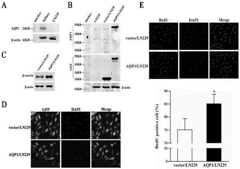
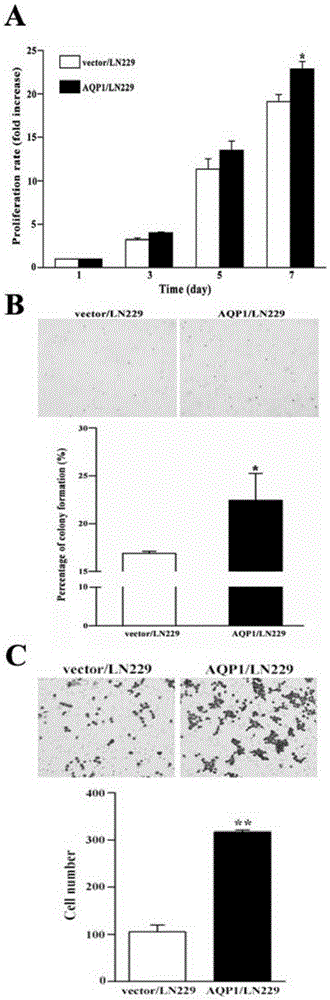
[0055] Overexpression of AQP1 improves the proliferation and invasion of LN229 cells
[0056] Previous studies have shown that AQP1 can enhance the cell migration ability of HT20, 12 / 40B16F10 and 4T1 cell lines (Jiang 2009, Hu&Verkman 2006). In order to study the role of AQP1 in glioma, we used Western Blot method to detect the expression of AQP1 in homologous LN229 cell line. AQP1 expression was not detected in LN229 cells ( figure 1 A). Then, we transfected the plasmid expressing the full-length AQP1 sequence into LN229 cells to obtain LN229 cells stably expressing AQP1, and named it AQP1 / LN229 cell line. The empty vector was transfected into LN229 cells to obtain LN229 cells stably expressing the empty vector, which were named vector / LN229 cell line as a control cell line. Compared with vehicle / LN229 cells, AQP1 protein expression level was significantly upregulated in AQP1 / LN229 cells ( figure 1 B). In order to complete the cellular localization of AQP1, an immunofluo...
Embodiment 2
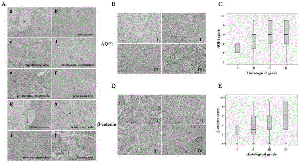
[0060] Expression of AQP1 in human glioma tissues
[0061]In order to further explore the functional role of AQP1 in glioma tissues, based on the gene expression analysis data from the Rembrandt database, we further detected the expression level of AQP1 mRNA. (Molecular Brain Tumor Data Lookup, https: / / caintegrator.nci.nih.gov / rembrandt / ). These data included glioma tissues (n=454) as well as non-tumor tissues (n=24). Valid data showed that AQP1mRNA levels were upregulated in glioma tissues (1.96±0.29) compared with non-tumor tissues ( image 3 A). Secondly, we used Western Blot to detect the expression level of AQP1 protein in various WHO grade gliomas ( image 3 B). Two normal brain samples and one nerve sheath sample were used as controls. It was found by Western Blot that the expression of AQP1 was not detected in the lysate of non-tumor tissue and schwannoma samples. In contrast, AQP1 expression levels were upregulated in human glioma tissues. Furthermore, AQP1 was...
Embodiment 3
[0063] Expression of AQP1 in patients with glioma
[0064] Interestingly, we observed that AQP1-immunohistochemistry showed specific patterns ( Figure 4 Aa-j). In contrast to previous studies, AQP1 expression was not detected in glial cells of normal brain tissue (Saadoun et al. 2002a, Hayashi et al. 2007). In contrast, AQP1 expression levels were upregulated in tumor regions ( Figure 4 Aa). In astrocytoma ( Figure 4 Ab) and oligodendroglioma ( Figure 4 Ac) AQP1 expression was observed. In astrocytomas, AQP1 staining presents a typical astrocyte appearance. In low-grade glioma endothelial cells, AQP1 expression was observed in human glioma tissue sections, whereas AQP1 expression was absent in proliferating endothelial cells of high-grade glioma. In addition, AQP1 immune responses were more aggregated and perivascularly distributed ( Figure 4 Af). Further observations revealed that AQP1 was expressed in tumor-infiltrated areas ( Figure 4 Ag) was expressed and l...
PUM
 Abstract
Description
Claims
Application Information
 - R&D
- Intellectual Property
- Life Sciences
- Materials
- Tech Scout
- Unparalleled Data Quality
- Higher Quality Content
- 60% Fewer Hallucinations
Browse by: Latest US Patents, China's latest patents, Technical Efficacy Thesaurus, Application Domain, Technology Topic, Popular Technical Reports.
© 2025 PatSnap. All rights reserved.Legal|Privacy policy|Modern Slavery Act Transparency Statement|Sitemap|About US| Contact US: help@patsnap.com