Device used for repeated immunostaining of same tissue slice

A technique for tissue sectioning and immunostaining, which is applied in the field of medical biological experimental devices, can solve the problems such as the difficulty of implementing the immunostaining method, and achieve the effect of facilitating repeated staining and photographing.

Pending Publication Date: 2017-01-04
NANCHANG DEMANDUO TECH CO LTD
View PDF5 Cites 5 Cited by
  • Summary
  • Abstract
  • Description
  • Claims
  • Application Information

AI Technical Summary

Problems solved by technology

If the study of the expression of multiple antigens is performed on a very small amount of tissue specimens (such as fine need

Method used


Image

  • Device used for repeated immunostaining of same tissue slice

Examples


Embodiment Construction

[0011] The present invention will be described in detail below in conjunction with the accompanying drawings and specific examples.

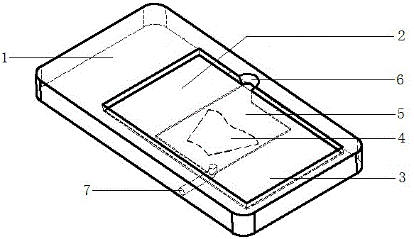
[0012] Such as figure 1 As shown, a device for performing multiple immunostaining on the same tissue section consists of a slide chamber 1 and a cover slip 3 .

[0013] The slide tank 1 is made of colorless transparent glass and has an upper groove 2 and a lower groove 5 that are continuous with each other. The size and shape of the upper groove 2 match the cover glass 3; the lower groove 5 is located at the center of the bottom of the upper groove 2, and the bottom of the lower groove 5 is flat, with a volume of 100 microliters. The cross-sectional area of ​​the lower groove 5 is preferably slightly larger than the area of ​​the tissue section 4. Near the right side wall, there is a liquid filling gap 6, and there is a liquid outlet hole 7 near the left side wall, which starts from the bottom of the lower groove 5 and passes out from the side ...


PUM


Abstract

In order to implement repeated immunostaining of a same tissue slice to save a tissue sample and meet the diagnosis requirement, the invention discloses a device for repeated immunostaining of the same tissue slice. The device is characterized by comprising a slide glass groove and a cover glass; the slide glass groove is provided with an upper groove and a lower groove which are mutually continued, and the size and the shape of the upper groove are matched with the size and the shape of the cover glass; the lower groove is located in the center of the bottom part of the upper groove, and the bottom part is flat; one face, facing downwards, of the cover glass is attached with the tissue slice; the tissue slice is located in a space formed by the cover glass and the lower groove. By using the device, the same tissue slice is subjected to repeated immunostaining and pictures are taken for the same tissue slice.

Description

technical field [0001] The invention relates to a medical biological experiment device, in particular to a device for performing multiple times of immunostaining on the same tissue section. Background technique [0002] Immunostaining is to use the principle of specific combination of antigen and antibody to detect the expression of specific protein (antigen). It is one of the most commonly used detection methods to discover the functional changes of the body under physiological or pathological conditions. The most commonly used method is immunohistochemical staining. and immunofluorescence staining. The conventional method is to stain a tissue section with one antigen marker, but to study the expression of multiple antigens requires multiple tissue sections. If a very small amount of tissue specimens (such as fine needle aspiration specimens) is used to study the expression of multiple antigens, conventional immunostaining methods are difficult to achieve due to the limita...

Claims


Application Information

Patent Timeline
IPC IPC(8): G01N1/31
CPCG01N1/312
Inventor 吴汉环其他发明人请求不公开姓名
Owner NANCHANG DEMANDUO TECH CO LTD
Why Eureka
Try Eureka