A left ventricular volume reduction device

A left ventricle and apex technology, applied in heart valve, medical science, surgery, etc., can solve the problems of limited shock absorption performance of the base, increased device cost, fatigue fracture, etc., to achieve enhanced volume reduction effect, enhanced blocking effect, strong The effect of fatigue resistance

Active Publication Date: 2019-01-25
NINGBO DIOCHANGE MEDICAL TECH CO LTD
View PDF4 Cites 0 Cited by
  • Summary
  • Abstract
  • Description
  • Claims
  • Application Information

AI Technical Summary

Problems solved by technology

This device has the following defects: 1. Clinically, the support rod of the support frame of the device, especially the far end of the support rod, should be pressed and held in the delivery catheter with a small inner diameter (about 4mm) in the delivery system, and then released on the left side. The ventricle is restored to a diameter of several tens of millimeters (65-95mm). Such a large amount of deformation leads to a support rod with a limited elastic deformation range, especially the proximal part of the support rod undergoes huge plastic deformation, so the support of the support frame The radial support force provided by the rod itself was seriously insufficient, and the device itself could not achieve adhesion to the wall of the left ventricle. Therefore, after the device was released, it still needed the assistance of a balloon catheter to expand, so that the device could adhere to the wall of the left ventricle, which resulted in a A series of problems, including: complex operation steps, prolonged operation time, which also increases the complexity of the delivery system structure; the increase of the balloon and the balloon channel in the delivery system increases the diameter of the delivery system, which in turn causes the device and the delivery system to enter the human blood vessel High risk of damage to blood vessels and heart valves when used with the heart; undoubtedly, it also increases the cost of the device and increases the economic burden on patients
2. According to clinical application, 12 months after operation, the left ventricular end-systolic volume (LVESV) decreased by 16% and the left ventricular end-diastolic volume (LVEDV) decreased by 14%. not enough
Although the left ventricular ejection fraction (LVEF) has increased moderately, the improvement of cardiac function including the pumping function of the left ventricle is not ideal, because the device basically does not have the auxiliary pumping function
3. After the device is released, because the blood in the heart generates a relatively large periodic impact force in the axial direction (parallel to the central axis of the support frame), the force impacts the center of the support rods of the device and then transfers to the base, and the device The supporting frame of the device does not have shock-absorbing performance, and the shock-absorbing performance of the base is very limited, which leads to stress concentration easily occurring on the supporting frame and the base of the device, and eventually fatigue fracture occurs. At present, relevant reports have been reported in the clinical follow-up of this product, which is also Can cause substantial damage to the apical part of the left ventricular wall by the device, such as puncturing the apex
4. After the support frame is released, the device can no longer be recovered, nor can it realize repeated positioning of the device

Method used


Image

  • A left ventricular volume reduction device
  • A left ventricular volume reduction device
  • A left ventricular volume reduction device

Examples


Embodiment 1

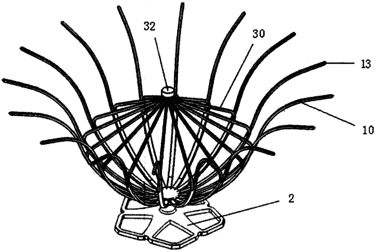
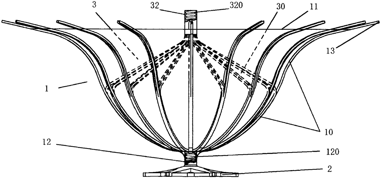
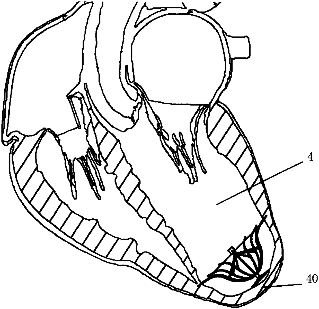
[0060] Such as figure 1 with 2 As shown, the left ventricular volume reduction device of the present invention includes a support frame 1 and a base 2 connected to the bottom 12 of the support frame 1. The support frame 1 is composed of a plurality of support rods 10, and the proximal ends of the support rods 10 are gathered to form a support frame 1, the distal end of the support rod 10 is adaptively opened in the direction of the distal end along the inner wall of the left ventricle 4, and the auxiliary umbrella 3 is also included in the space defined by the plurality of support rods 10, the auxiliary umbrella 3 is composed of a plurality of auxiliary umbrella ribs 30, the auxiliary ribs 30 extend from the central end 32 to the apex 40, and their terminals are connected to the middle of the support rods 10. Multiple auxiliary ribs 30 are in one-to-one correspondence with multiple support rods 10, and the surface of the support frame 1 is all Covered with a first film 11 . ...

Embodiment 2

[0067] Such as Figure 5 As shown, based on Embodiment 1, the difference between Embodiment 2 and Embodiment 1 is that the surface of the support frame 1 is only partially covered with the first film 11, that is, the support rod 10 and the auxiliary umbrella on the support frame 1 are covered. The connection point of the bone 30 extends to the distal area of ​​the support frame 1, while the surface of the auxiliary umbrella 3 is entirely covered with the second membrane 31, and the proximal end of the first membrane 11 is in a sealed connection with the proximal end of the second membrane 31.

[0068] The optional membrane material of the second membrane 31 on the auxiliary umbrella 3 includes expanded polytetrafluoroethylene (ePTFE), polyester (PET), polyurethane elastomer (TPU), polyamide (PA), silica gel, degradable materials such as Polylactic acid (PLA), animal tissue, etc., after the device is released, due to the introduction of the second membrane 31, the device exhibi...

Embodiment 3

[0071] Such as Figure 7 As shown, based on Embodiment 1, the difference between Embodiment 3 and Embodiment 1 is that the proximal end of the auxiliary rib 30 of the auxiliary umbrella 3 is connected to the bottom 12, and the surface of the auxiliary umbrella 3 is completely covered with a second film 31. A plurality of auxiliary umbrella ribs 30 and the second polymer film 18 define a bladder 33, and the bladder 33 is preferably spherical or ellipsoidal. Here, the number of the auxiliary ribs 30 and the support rods 10 can be consistent or inconsistent. When the number of the auxiliary ribs 30 and the support rods 10 are inconsistent, only the auxiliary ribs 30 and the second membrane 31 can form a capsule 33. It is enough to increase the shock absorption effect and improve the durable life of the device, especially the bladder 33.

[0072] The proximal end of the auxiliary umbrella rib 30 is connected to the bottom 12 of the support frame 1, so that the device can support ...


PUM


Abstract

The invention relates to a left ventricular volume reduction device, which includes a support frame and a base connected to the bottom of the support frame. The support frame is composed of a plurality of support rods. The proximal ends of the support rods are gathered to form the bottom of the support frame. The distal ends of the support rods are It opens adaptively along the inner wall of the left ventricle in the direction away from the apex, and also includes an auxiliary umbrella in the space defined by multiple support rods. The auxiliary umbrella is composed of multiple auxiliary ribs, and the auxiliary ribs extend from the central end toward the apex. The terminal is connected to the support rod, the surface of the support frame is covered with film, and multiple auxiliary ribs radially support the multiple support rods so that the support frame adapts to the inner wall of the left ventricle. The invention solves the problems in the prior art that the structure of the delivery system is complex and the surgical steps are complicated; the volume reduction effect is good, the ventricular pumping function is enhanced, the blocking effect is enhanced, the operation is simple, and the positioning and recovery can be repeated.

Description

technical field [0001] The invention relates to the technical field of medical ventricular isolation devices, in particular to a left ventricular volume reduction device. Background technique [0002] In recent years, the prevalence of heart failure has increased year by year and has become a serious public health problem. According to reports, 17 million people die of cardiovascular disease in the world every year, and more than half of them die of acute myocardial infarction. Even if patients survive, they are prone to develop heart failure. chronic heart failure. It is estimated that the incidence of acute myocardial infarction in China is about 45 / 100,000 to 55 / 100,000, and it is still on the rise. The prevalence of heart failure is 0.9%, and the cause of coronary heart disease has increased from 36.8% in 1980 to 2000. 45.6%, ranking first in various etiologies. About 20% to 50% of patients after acute myocardial infarction will develop heart failure, especially anteri...

Claims


Application Information

Patent Timeline
Patent Type & Authority Patents(China)
IPC IPC(8): A61B17/00A61B17/12A61F2/24
CPCA61B17/0057A61B17/12022A61B17/12122A61B17/1215A61F2/2487A61B17/1214A61B2017/12095A61B2017/00243A61B2017/00615A61B2017/00592A61B2017/00575A61B2017/00867A61B2017/00632
Inventor 李彪葛均波吕世文周达新李欣欣沈雳
Owner NINGBO DIOCHANGE MEDICAL TECH CO LTD
Why Eureka
Try Eureka