Device for modifying an imaging of a tee probe in X-ray data

An X-ray and data technology, applied in the direction of radiological diagnostic equipment, image data processing, application, etc., can solve the problems of time-consuming, clinical workflow disruption, etc., to reduce risks, improve clinical workflow, and easy to control Effect

Active Publication Date: 2017-02-22
KONINKLJIJKE PHILIPS NV
View PDF10 Cites 0 Cited by
  • Summary
  • Abstract
  • Description
  • Claims
  • Application Information

AI Technical Summary

Problems solved by technology

[0009] This entire process is disruptive to clinical workflow and time consuming for both interventional cardiologists and echocardiologists
It is also more risky for the patient as this extra movement and adjustment of the TEE probe within the esophagus can put the patient at some additional risk

Method used


Image

  • Device for modifying an imaging of a tee probe in X-ray data
  • Device for modifying an imaging of a tee probe in X-ray data
  • Device for modifying an imaging of a tee probe in X-ray data

Examples


Embodiment Construction

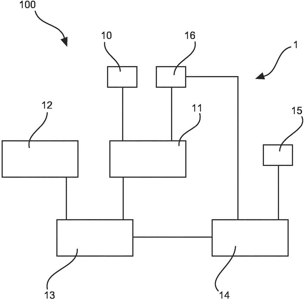
[0046] figure 1 A schematic diagram of an example of a medical imaging system 100 for modifying imaging of a TEE probe in X-ray data is shown. The medical imaging system 100 comprises a device 1 for modifying imaging of a TEE probe in X-ray data, a TEE probe and an image acquisition device 102 .

[0047] The device includes an X-ray data providing unit 11 , a model providing unit 12 , a position locating unit 13 and a processing unit 14 . The X-ray data providing unit 11 provides X-ray image data including image data of the TEE probe and is thus connected to the image acquisition device 102 . The model providing unit 12 provides model data of the TEE probe. The model data may be a 3D model of the TEE probe. The 3D model of the TEE probe can be a 3D CAD model, a 3D model of the X-ray attenuation of the TEE probe (eg from a CT scan), etc.

[0048] The position locating unit 13 locates the position of the TEE probe in the X-ray data based on the model data of the TEE probe. ...


PUM


Abstract

The present invention relates to a device (1) for modifying an imaging of a TEE probe in X-ray data, a medical imaging system (100) for modifying an imaging of a TEE probe in X-ray data, a method for modifying an imaging of a TEE probe in X-ray data, a computer program element for controlling such device (1) and a computer readable medium having stored such computer program element. The device (1) comprises an X-ray data provision unit (11), a model provision unit (12), a position locating unit (13), and a processing unit (14). The X-ray data provision unit (11) is configured to provide X-ray data comprising image data of a TEE probe. The model provision unit (12) is configured to provide model data of the TEE probe. The position locating unit (13) is configured to locate a position of the TEE probe in the X-ray data based on the model data of the TEE probe. The processing unit (14) is configured to define a region in a predetermined range adjacent to the TEE probe as reference area. The processing unit (14) is configured to process the X-ray data of the reference area into estimated X-ray data of a region occupied by the TEE probe. The processing unit (14) is configured to modify the X-ray data in the region occupied by the TEE probe based on the estimated X-ray data.

Description

technical field [0001] The invention relates to a device for modifying the imaging of a TEE probe in X-ray data, a medical imaging system for modifying the imaging of a TEE probe in X-ray data, and a device for modifying the imaging of a TEE probe in X-ray data. A method of imaging a TEE probe, a computer program element for controlling such a device, and a computer-readable storage medium storing such a computer program element. Background technique [0002] X-ray imaging (fluoroscopy and angiography) is an important imaging modality for cardiac interventions. To provide guidance for procedures that require more soft tissue information than X-ray imaging provides, transesophageal echocardiography (TEE) information is employed. TEE imaging, which is a form of ultrasound imaging, is capable of showing, for example, an interventional device and its surrounding anatomy simultaneously, and provides fine detail of anatomy and function, which is often missing in live X-ray images...

Claims


Application Information

Patent Timeline
Patent Type & Authority Applications(China)
IPC IPC(8): A61B6/00A61B6/12A61B8/12A61B8/00
CPCA61B6/12A61B6/463A61B6/467A61B6/5235A61B8/12A61B8/445A61B6/503A61B6/5252A61B6/5258G06T5/008G06T2207/10116G06V2201/034A61B6/4441A61B6/461G06T7/0012G06T2207/30048
Inventor J·A·布拉肯N·奈霍夫M·格拉斯
Owner KONINKLJIJKE PHILIPS NV
Why Eureka
Try Eureka