Convolutional neural network-based kidney tubule epithelial cell automatic detection method

A convolutional neural network and epithelial cell technology, applied in the field of automatic detection of renal tubular epithelial cells, can solve problems such as easy fatigue, missed detection, and false detection, and achieve the effect of avoiding low efficiency

Inactive Publication Date: 2017-03-08
UNIV OF ELECTRONIC SCI & TECH OF CHINA
View PDF4 Cites 8 Cited by
  • Summary
  • Abstract
  • Description
  • Claims
  • Application Information

AI Technical Summary

Problems solved by technology

Since the detection of renal tubular epithelial cells has great medical reference significance for the localization and diagnosis of renal parenchymal diseases in patients, however, most hosp

Method used


Image

  • Convolutional neural network-based kidney tubule epithelial cell automatic detection method
  • Convolutional neural network-based kidney tubule epithelial cell automatic detection method

Examples


Embodiment Construction

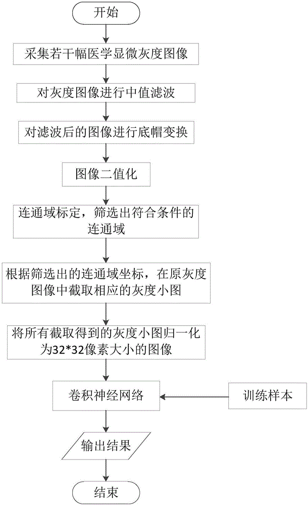
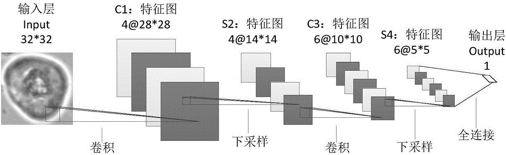
[0034] Below in conjunction with accompanying drawing, a kind of automatic detection method of renal tubular epithelial cell based on convolutional neural network of the present invention is described in detail:

[0035] Step 1: Use a microscope to collect several medical microscopic grayscale images;

[0036] Step 2: Perform median filtering on the obtained grayscale image;

[0037] Step 2-1: Slide the 3×3 filter window as a template on the original image, and the center position of the filter window is the target pixel to be processed by the filter;

[0038] Step 2-2: Sort the pixel gray values ​​in the filtering window according to their size to obtain the median value, and assign the obtained median value to the original target pixel;

[0039] Step 2-3: After the target pixel gets a new gray value, check whether the image has been traversed and calculated. If there are still pixels in the image that have not been calculated, return to continue sliding the filter window to...


PUM


Abstract

The invention discloses a convolutional neural network-based kidney tubule epithelial cell automatic detection method and belongs to the image processing field. According to the convolutional neural network-based kidney tubule epithelial cell automatic detection method, cells are screened initially based on the morphological features of kidney tubule epithelial cells; identification is carried out based on a convolutional neural network; and therefore, automatic detection of the kidney tubule epithelial cells can be realized, and defects such as low detection efficiency and susceptibility to subjective factors can be avoided, and efficient and convenient detection can be realized. The number of visible components in medical microscopic images is large, and the backgrounds of the medical microscopic images are complex, and as a result, medical personnel tend to generate visual fatigue in a long-term high-intensity working condition, and the possibility of misjudgment or omitted judgment in microscopic examination is large. According to the convolutional neural network-based kidney tubule epithelial cell automatic detection method of the invention, the automatic detection of the kidney tubule epithelial cells can be realized based on digital image processing technologies, and therefore, detection accuracy can be improved, the risk of treatment delay of patients can be lowered, and the labor intensity of the medical personnel can be reduced.

Description

technical field [0001] The invention belongs to the technical field of biomedical image processing, and in particular relates to an automatic detection method of renal tubular epithelial cells based on a convolutional neural network. Background technique [0002] Renal tubular epithelial cells are a formed component in urine, and the content in normal people's urine is zero or very little. However, for patients with renal tubular lesions, a large number of renal tubular epithelial cells can be found in urine sediment examination, gynecological leucorrhea routine examination (for women), and semen examination (for men). Since the detection of renal tubular epithelial cells has great medical reference significance for the localization and diagnosis of renal parenchymal diseases in patients, however, most hospitals now use manual microscopic examination for the detection of the cells. For reasons such as high-intensity work for a long time and easy fatigue, false detections an...

Claims


Application Information

Patent Timeline
IPC IPC(8): G06T7/00G06K9/00
CPCG06T7/0012G06T2207/20081G06T2207/30084G06T2207/10056G06V20/695
Inventor 张静郝如茜陈祥韩翠张正龙胡静蓉杜晓辉刘娟秀倪光明刘霖刘永
Owner UNIV OF ELECTRONIC SCI & TECH OF CHINA
Why Eureka
Try Eureka