
Lung tissue segmentation method, apparatus, medical image processing system

A medical image and lung tissue technology, applied in the field of medical image processing, can solve problems such as the inability to achieve tissue and organ segmentation, and achieve the effect of eliminating influence

Active Publication Date: 2017-04-05
SHANGHAI UNITED IMAGING HEALTHCARE
View PDF7 Cites 17 Cited by
  • Summary
  • Abstract
  • Description
  • Claims
  • Application Information

AI Technical Summary

Problems solved by technology

In the existing technology, the accuracy of the method based on region segmentation needs to be further improved, and the segmentation of different tissues and organs in multi-bed MRI images cannot be realized

Method used


Image

  • Lung tissue segmentation method, apparatus, medical image processing system
  • Lung tissue segmentation method, apparatus, medical image processing system
  • Lung tissue segmentation method, apparatus, medical image processing system

Examples


Embodiment Construction

[0055]In order to more clearly illustrate the technical solutions of the embodiments of the present application, the following briefly introduces the drawings that need to be used in the description of the embodiments. Obviously, the accompanying drawings in the following description are only some examples or embodiments of the present application, and those skilled in the art can also apply the present application to other similar scenarios. Unless otherwise apparent from context or otherwise indicated, like reference numerals in the figures represent like structures or operations.

[0056] As indicated in this application and claims, the terms "a", "an", "an" and / or "the" do not refer to the singular and may include the plural unless the context clearly indicates an exception. Generally speaking, the terms "comprising" and "comprising" only suggest the inclusion of clearly identified steps and elements, and these steps and elements do not constitute an exclusive list, and t...


PUM


Abstract

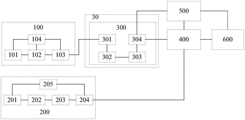
The invention discloses a ling tissue segmentation method, comprising: obtaining a first medical image of the target area of an examinee wherein the first medical image consists of a plurality of layered images with each layered image containing a plurality if pixels; linearly enhancing the sagittal plane of the first medical image; adding the gray values of the pixels corresponding to the first medical image and the linearly enhanced first image to generate a second medical image; determining the sagittal skin line of the second medical image and determining the cross-sectional skin line of the second medical image on the sagittal skin line determined second medical image; obtaining the connected domain enclosed by the sagittal skin line and the cross-sectional skin line; and obtaining the lung tissue segmentation result in the connected domain. The lung tissue segmentation method of the invention can accurately obtain the boundary area of a lung tissue. At the same time, the invention also proposes lung tissue segmentation apparatus and a medical image processing system using the lung tissue segmentation apparatus.

Description

technical field [0001] The invention relates to the technical field of medical image processing, in particular to a lung tissue segmentation method, device and medical image processing system in medical images. Background technique [0002] Emission computed tomography (ECT) mainly includes single photon emission computed tomography (SPECT) and positron emission computed tomography (PET). It is an indispensable and important method for the diagnosis of heart and brain diseases. Magnetic resonance imaging (Magnetic Resonance Imaging, MRI) can provide information on the anatomical shape and physiological function of the imaging site, and has incomparable advantages in reflecting anatomical shape and physiological function information, especially soft tissue MR images present better contrast effects , and no radiation. The combination of PET and MR examination has the advantages of good sensitivity and high accuracy. In the PET / MR multimodal imaging system, attenuation corre...

Claims


Application Information

Patent Timeline
IPC IPC(8): G06T7/00G06T7/11G06T7/187G06T7/194
CPCG06T7/0012G06T2207/10088G06T2207/10104G06T2207/30061
Inventor 李鹏程姜娈李强
Owner SHANGHAI UNITED IMAGING HEALTHCARE
Learn More