Method for detecting entry of small molecule compounds into mitochondria at cell in-situ level

A technology of small molecule compounds and mitochondria, which is applied in the field of molecular pharmacology, can solve the problems that cannot explain the way small molecule drugs enter the mitochondria, and achieve the effect of no background interference, high sensitivity and high sensitivity

Active Publication Date: 2017-05-31
江西海普洛斯医学检验实验室有限公司
View PDF5 Cites 2 Cited by
  • Summary
  • Abstract
  • Description
  • Claims
  • Application Information

AI Technical Summary

Problems solved by technology

However, the use of this type of detection method has great limitations. First, the compound is required to have a fluorescent group, which is not applicable to most compounds; secondly, this type of detectio...

Method used


Image

  • Method for detecting entry of small molecule compounds into mitochondria at cell in-situ level
  • Method for detecting entry of small molecule compounds into mitochondria at cell in-situ level
  • Method for detecting entry of small molecule compounds into mitochondria at cell in-situ level

Examples


Embodiment 1

[0038] Embodiment 1: the preparation of the small molecule compound (hereinafter represented by NA17) of structure shown in formula (A)

[0039] The synthetic route of NA17 is shown below, and the reagents and conditions are as follows: a: piperethylamine, ethanol, 70°C, stirred for 10 hours; b: propylamine, DMSO, 135°C:

[0040]

[0041] The compound (2.77g, 10mmol) of the structure shown in the formula (B) was dissolved in 150ml ethanol (100v / v%), fully stirred, then added piperonyl ethylamine (1.65g, 10mmol), and the reaction mixture was heated at 70°C The reaction was stirred for 10 hours. After the reaction was over, the solution was cooled to room temperature and filtered, and the filter cake was collected to obtain an intermediate product of the structure shown in formula (C), which was directly used in the next step without further purification; (C) The intermediate product (4.23g, 10mmol) and 3-dimethylaminopropylamine (1.02g, 10mmol) of the structure shown in (C) ...

Embodiment 2

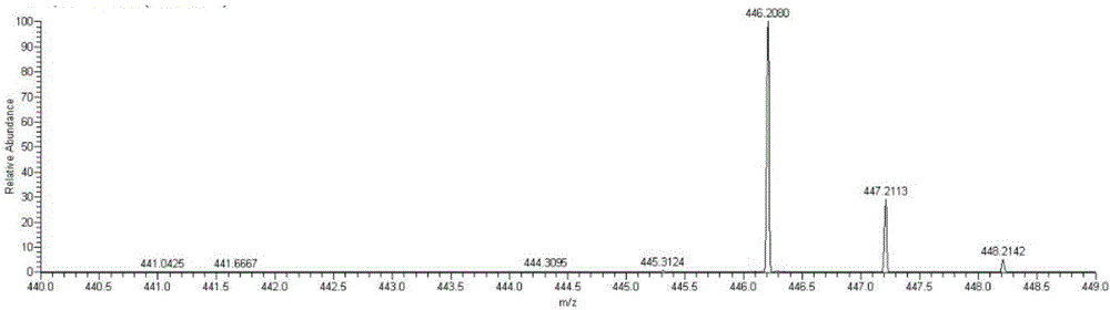
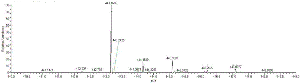
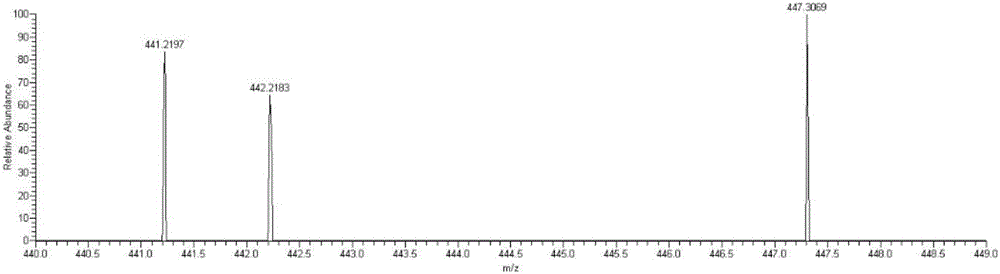
[0043] Example 2: Detecting whether NA17 can enter mitochondria at the in situ level of cells

[0044] (1) Cell administration

[0045] 1. Inoculate the non-small cell lung cancer cell line NCI-H460 on a 70cm culture dish, carefully remove the medium in the culture dish when the cell confluence reaches 50% to 60%, wash twice with PBS, and add new DMEM culture medium .

[0046] 2. 2 mM NA17 original drug was prepared with DMSO as a solvent, added to a petri dish so that the final concentration was 10 μM, administered for 5 hours, and a control group without drug addition was set at the same time.

[0047] (2) Separation of mitochondria

[0048] 1. Carefully remove the medium in the culture dish after the administration treatment, wash twice with PBS, digest the adherent cells with 0.25% trypsin; add 5mL of PBS solution, blow off the adherent cells and collect the suspension in a 10mL centrifuge centrifuge at 2000rpm for 10min in a centrifuge;

[0049] 2. Discard the superna...


PUM


Abstract

The invention discloses a method for detecting entry of small molecule compounds into mitochondria at a cell in-situ level. The method particularly comprises the following steps: selecting suitable small molecule compounds to be incubated with cells; acquiring mitochondria with complete physiological structures without destroying cell mitochondria; collecting cytoplasm; incubating with the small molecule compounds and the acquired mitochondria; respectively pyrolyzing the obtained mitochondria after incubating the small molecule compounds and the cytoplasm collected before under non-denaturing conditions to acquire a mitochondrial protein lysate and a cytoplasmic protein lysate; and then, detecting whether the small molecule compounds enter the mitochondria at the cell in-situ level by combining with mass spectrum analysis. The method can be adopted to detect whether the small molecule compounds enter the mitochondria at the cell in-situ level, and can further detect a way in which the small molecule compounds enter the mitochondria, can be directly applied to target region verification of a pilot drug or screening of the pilot drug using the mitochondrion as an effector, and has the advantages of high reliability and high sensitivity.

Description

technical field [0001] The invention relates to the field of molecular pharmacology, in particular to a method for in situ detection of small molecule compounds entering mitochondria in cells. Background technique [0002] As a key organelle in cells, mitochondria not only have important functions such as supplying energy for cells and participating in metabolism, but also participate in important biological processes such as cell signal transduction and cell apoptosis. A large number of studies have shown that the number, distribution, structure and function of mitochondria are closely related to neurodegenerative diseases (such as Parkinson's disease, Alzheimer's disease), metabolic diseases (such as type II diabetes, obesity), cancer and cardiovascular diseases diseases are closely related. Mitochondria are dubbed as "cell signal transduction organelle" and "motor of cell death", and the related "mitochondria" has become a research hotspot in the fields of life science a...

Claims


Application Information

Patent Timeline
IPC IPC(8): G01N27/62
CPCG01N27/62
Inventor 张国海彭艳杨阳李亮萍曾淑兰
Owner 江西海普洛斯医学检验实验室有限公司
Why Eureka
Try Eureka