MRI (magnetic resonance imaging) method and device

A technology of magnetic resonance imaging and imaging method, which is applied in the directions of measuring devices, measuring magnetic variables, instruments, etc., can solve the problems of long scanning time, etc., and achieve the effect of high signal-to-noise ratio, high imaging resolution, and improved imaging speed

Active Publication Date: 2017-05-31
SHENZHEN INST OF ADVANCED TECH
View PDF6 Cites 7 Cited by
  • Summary
  • Abstract
  • Description
  • Claims
  • Application Information

AI Technical Summary

Problems solved by technology

[0004] In view of this, the embodiment of the present invention provides a magnetic re...

Method used


Image

  • MRI (magnetic resonance imaging) method and device
  • MRI (magnetic resonance imaging) method and device
  • MRI (magnetic resonance imaging) method and device

Examples


Embodiment 1

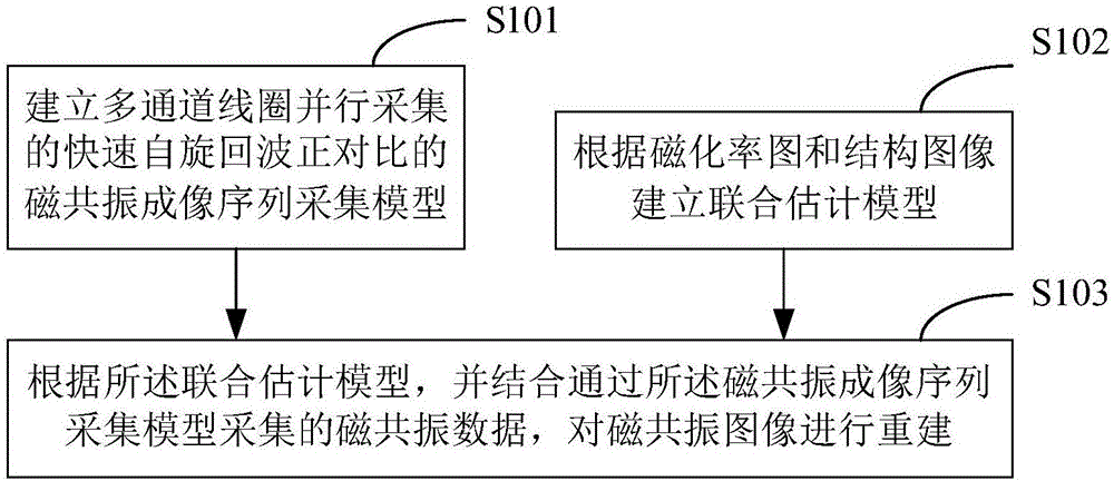
[0047] figure 1 The implementation process of the magnetic resonance imaging method provided by Embodiment 1 of the present invention is shown, and the details are as follows:

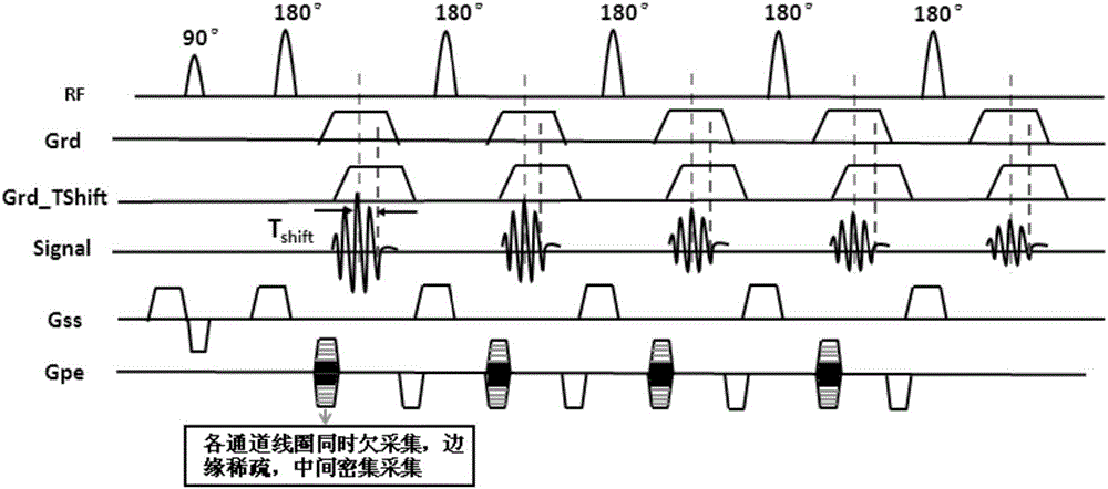
[0048] Step S101 , establishing an MRI sequence acquisition model for fast spin-echo direct contrast acquired by multi-channel coils in parallel.

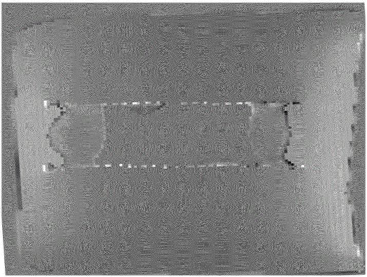
[0049] It can be understood that since the magnetically compatible metal interventional devices do not have H protons, they show signal loss in their own position areas on traditional MR imaging images, and these metal interventional devices will be magnetized in the MR external magnetic field, resulting in The local magnetic field, which interferes with the surrounding tissue region, i.e. susceptibility artifacts, thus exhibits a large black hole (the region is much larger than the size of the device itself) in the area around the interventional device, i.e. negative contrast Image, the negative contrast of the image makes it very difficult to distinguish...

Embodiment 2

[0079] Corresponding to the magnetic resonance imaging method described in the first embodiment above, Figure 9 A structural block diagram of the magnetic resonance imaging apparatus provided by Embodiment 2 of the present invention is shown. For ease of description, only the parts related to this embodiment are shown.

[0080] refer to Figure 9 , the device includes a first model building module 101 , a second model building module 102 and a reconstruction module 103 . Wherein, the first model building module 101 is used for building a fast spin echo positive contrast magnetic resonance imaging sequence acquisition model for parallel acquisition by multi-channel coils. The second model building module 102 is used for building a joint estimation model according to the magnetic susceptibility map and the structure image. The reconstruction module 103 is configured to reconstruct the magnetic resonance image according to the joint estimation model combined with the acquisit...


PUM


Abstract

The invention applies to the technical field of magnetic resonance and provides an MRI (magnetic resonance imaging) method and device. The MRI method comprises the following steps: establishing an MRI sequence acquisition model for positive contrast of FSE (Fast spin echo) acquired parallelly by multiple-channel coils; establishing a joint estimation model according to a magnetic susceptibility map and a structure image; acquiring MRI data according to the joint estimation model in combination with the MRI sequence acquisition model so as to reconstruct an MRI image. With adoption of the method, the positional relation between an intervention device and surrounding tissue can be recognized clearly, intraductal condition of a stent device is evaluated, the imaging resolution is higher, the imaging speed is higher, and the signal-to-noise ratio is higher.

Description

technical field [0001] The invention belongs to the technical field of magnetic resonance, and in particular relates to a magnetic resonance imaging method and device. Background technique [0002] MRI (Magnetic Resonance Imaging, Magnetic Resonance Imaging) has excellent soft tissue contrast, can be multi-parameter, multi-plane imaging at any angle, has high sensitivity to flowing tissue, and more importantly, has many advantages such as non-invasive, no X-ray radiation damage, etc. , making it widely used in clinical diagnosis and medical research. MRI is based on the H protons that are abundant in human tissues, and a resonance signal is generated by adding a radio frequency pulse with the same frequency as the H proton spin. Since the magnetically compatible metal interventional device does not have H protons, it shows signal loss in its own position area on the traditional MR imaging image, and it is difficult to identify the characteristics of the surrounding tissue o...

Claims


Application Information

Patent Timeline
IPC IPC(8): G01R33/561
CPCG01R33/5611
Inventor 史彩云谢国喜苏适张晓咏纪秀全刘新
Owner SHENZHEN INST OF ADVANCED TECH
Why Eureka
Try Eureka