FPGA-based spatial composite imaging method and device

A technology of spatial compounding and imaging method, applied in the field of medical ultrasound imaging, can solve the problems of slow processing speed and low imaging frame rate, and achieve the effect of improving speed, improving imaging frame rate, and reducing processing time

Active Publication Date: 2020-02-14
SONOSCAPE MEDICAL CORP
View PDF4 Cites 0 Cited by
  • Summary
  • Abstract
  • Description
  • Claims
  • Application Information

AI Technical Summary

Problems solved by technology

[0005] The object of the present invention is to provide a spatial composite imaging method and device to solve the problems of slow processing speed and low imaging frame rate of the existing spatial composite

Method used


Image

  • FPGA-based spatial composite imaging method and device
  • FPGA-based spatial composite imaging method and device
  • FPGA-based spatial composite imaging method and device

Examples


Embodiment Construction

[0054] In order to enable those skilled in the art to better understand the solution of the present invention, the present invention will be further described in detail below in conjunction with the accompanying drawings and specific embodiments. Apparently, the described embodiments are only some of the embodiments of the present invention, but not all of them. Based on the embodiments of the present invention, all other embodiments obtained by persons of ordinary skill in the art without making creative efforts belong to the protection scope of the present invention.

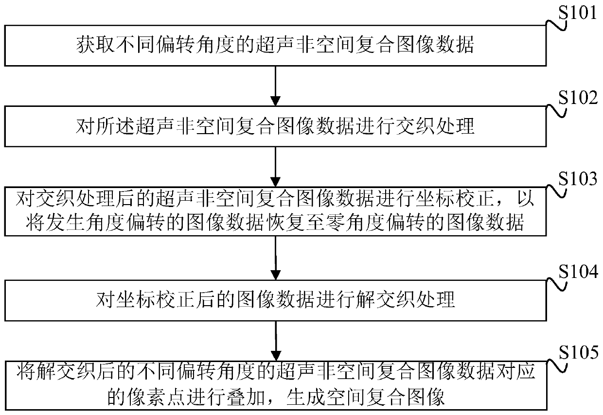
[0055] A flow chart of a specific embodiment of the FPGA-based spatial compound imaging method provided by the present invention, as figure 1 As shown, the method includes:

[0056] Step S101: Obtain ultrasonic non-spatial composite image data at different deflection angles.

[0057] The ultrasonic probe emits ultrasonic signals and receives ultrasonic echo signals with different deflection angles. In addition...


PUM


Abstract

An FPGA-based space compound imaging method and device. The method comprises: acquiring ultrasonic non-space compound image data at different deflection angles (S101); carrying out interleaving processing on the ultrasonic non-space compound image data (S102); correcting the coordinates of the ultrasonic non-space compound image data, which has undergone interleaving processing, so as to recover image data subjected to angle deflection to image data with zero angle deflection (S103); carrying out de-interleaving processing on the image data, the coordinates thereof having been corrected (S104); and superposing pixel points corresponding to the de-interleaved ultrasonic non-space compound image data with different deflection angles, and generating a space compound image (S105). The present invention reduces the complexity of implementing a space compound algorithm, can effectively improve a space compound calculation speed, reduces the processing time for an image, and improves an imaging frame rate.

Description

technical field [0001] The invention relates to the technical field of medical ultrasonic imaging, in particular to an FPGA-based spatial compound imaging method and device. Background technique [0002] The spatial composite imaging in the medical ultrasound imaging system is an imaging method that scans the scanning object along different angles, and then superimposes the corresponding pixel points of the images of these different angles to form an image. Spatial compound imaging technology can improve the detailed performance of images, reduce the influence of speckle noise, clutter and other ultrasonic artifacts on image quality, enhance the resolution ability between tissues, and significantly improve the low-contrast tissues and tissues in ultrasonic images. The definition of small lesions clearly shows the boundaries between tissues. [0003] The existing spatial compound imaging technology scans the target tissue from different angles through the deflection of the s...

Claims


Application Information

Patent Timeline
Patent Type & Authority Patents(China)
IPC IPC(8): G06T3/60G06T5/50G06T7/10G06T5/00G06T9/00
CPCG06T3/60G06T5/002G06T5/50G06T9/00G06T2207/10132G06T2207/30004
Inventor 朱曦叶宗英刘辉黄明军
Owner SONOSCAPE MEDICAL CORP
Why Eureka
Try Eureka