
A computer-based device and method for detecting eye movement distance and binocular movement consistency deviation

An eye movement and computer technology, applied in the field of medical devices, can solve the problems of inability to accurately detect eye movement distance and eye movement distance, and achieve the effects of low cost, easy operation, accurate detection and quantification

Active Publication Date: 2019-03-15
周凌云 +1
View PDF4 Cites 0 Cited by
  • Summary
  • Abstract
  • Description
  • Claims
  • Application Information

AI Technical Summary

Problems solved by technology

[0005] The purpose of the present invention is to solve the problem that the existing method cannot detect the eyeball movement distance quickly and accurately, and the eyeball movement distance cannot be detected when the eyeball is blocked, and provides a computer-based eye movement distance and binocular movement consistency deviation detection Device and method

Method used


Image

  • A computer-based device and method for detecting eye movement distance and binocular movement consistency deviation
  • A computer-based device and method for detecting eye movement distance and binocular movement consistency deviation

Examples


specific Embodiment approach 1

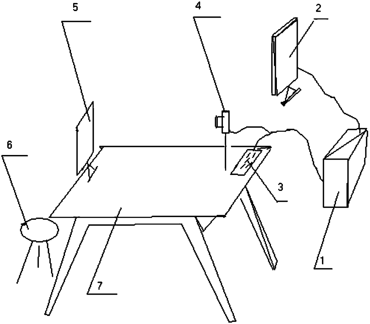
[0030] Specific implementation mode one: combine figure 1 Describe this embodiment, a computer-based eye movement distance and binocular movement consistency deviation detection device described in this embodiment includes a head fixing mechanism 5, a camera 4 and a computer;

[0031] The head fixing mechanism 5 is used to fix the patient's head, and the camera 4 is used to display the patient's binocular images in real time and send the images to the computer;

[0032] The detection module realized by software is embedded in the computer, and the software adopts VisualC++6.0 to write, and this module includes the following units:

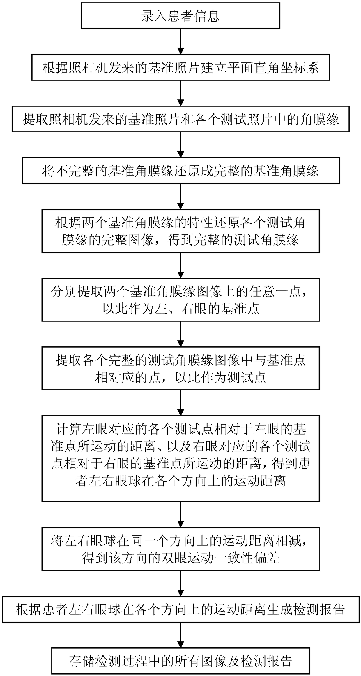
[0033] Coordinate system establishing unit: establish a plane Cartesian coordinate system according to the reference photo sent by the camera 4, the reference photo refers to the photo taken when the patient looks directly at the camera 4;

[0034] Limbal extraction unit: extract the limbus from the reference photos sent by the camera 4 and the li...

specific Embodiment approach 2

[0069] Specific Embodiment 2: This embodiment is a further limitation of the detection device described in Embodiment 1. In this embodiment, the detection module further includes:

[0070] Reference limbus restoration unit: restore the incomplete reference limbus to a complete reference limbus;

[0071] The reference point extracting unit extracts reference points from the complete reference limbus image.

[0072] The size of the eye fissure varies from person to person. People with large palpebral fissures can capture the entire limbus when taking baseline photos, but people with small palpebral fissures can only capture most of the limbus. For this reason, in this embodiment, a reference limbus restoration unit is added to restore the complete reference limbus to obtain a complete reference limbus image. It is more convenient to use the complete reference limbus image to select the reference point.

specific Embodiment approach 3

[0073] Specific Embodiment 3: This embodiment is a further limitation of the detection device described in Embodiments 1 and 2. In this embodiment, the method for extracting the limbus by the limbus extraction unit is: using computer image recognition technology, A test photo is compared with the reference photo, and the same part in the two photos is removed to obtain the reference limbus and the test limbus.

[0074] In this embodiment, the difference image method is used to extract the limbus. Using computer image recognition technology, any test photo is compared with the reference photo, and the same part in the two photos is removed, so that only two limbus remain in each photo.


PUM


Abstract

The invention discloses an eyeball movement distance and binocular movement consistency deviation detecting device and method based on a computer, relates to the field of medical instruments and aims to solve the problem that existing methods cannot quickly and accurately detect eyeball movement distance and cannot detect the eyeball movement distance when eyeballs are blocked. A head fixing mechanism is used for fixing the head of a patient, and a camera is used for displaying binocular images of the patient in real time and sending the images to the computer; the computer builds a plane rectangular coordinate system according to a reference image, extracts corneal limbi in the reference image and each test picture, reduces a complete image of each test corneal limbus according to characteristics of two reference corneal limbi, respectively extracts reference points on two reference corneal limbus images and test points corresponding to the reference points in test corneal limbus images and finally calculates distances by which each test point moves relative to the reference points and binocular movement consistency deviation. The eyeball movement distance and binocular movement consistency deviation detecting device and method is suitable for assessing eyeball movement functions.

Description

technical field [0001] The invention relates to the field of medical equipment, in particular to the detection of eyeball movement function. Background technique [0002] The left and right eyes of normal people can flexibly rotate in all directions. When the eyeball moves to the limit position and keeps gazing, both eyes can reach the same position to ensure that the visual object is a single image. When ocular motor nerve paralysis or extraocular muscle disease When it occurs, there is incoordination of binocular movement, which is manifested by the inability of the eyeballs to move in one direction or in multiple directions, which is also called eye movement disorder. Eye movement disorders are caused by damage to the cranial nerves due to various causes. Due to the different causes and the severity of the disease, the degree of movement disorders varies, and the condition of eye movement disorders will change during the development and outcome of the disease. The condit...

Claims


Application Information

Patent Timeline
Patent Type & Authority Patents(China)
IPC IPC(8): A61B3/113A61B3/14
CPCA61B3/0016A61B3/0083A61B3/113A61B3/14
Inventor 周凌云周晋刘铁镌栗雪梅
Owner 周凌云
Learn More