CT image pulmonary parenchyma template weasand elimination method based on breadth-first search

A breadth-first search and CT image technology, applied in image enhancement, image analysis, image data processing, etc., can solve problems such as interference and affecting the accuracy of recognition, and achieve the goals of reducing errors, improving efficiency, and increasing workload and complexity Effect

Active Publication Date: 2017-06-20
ZHEJIANG UNIV
View PDF8 Cites 6 Cited by
  • Summary
  • Abstract
  • Description
  • Claims
  • Application Information

AI Technical Summary

Problems solved by technology

In computer-aided detection, it is necessary to perform feature extraction, analysis, and recognition on lung CT images. In addition to lung parenchyma, there are some other organs and tissues in ordinary lung CT images, as well as the background of CT images. The feature extraction of a CT image will affect the accuracy of recognition. Therefore, it is necessary to segment the lung parenchyma on the CT image of the lungs.
In the existing methods, the lung parenchyma segmentation template is often obtained by iterative

Method used


Image

  • CT image pulmonary parenchyma template weasand elimination method based on breadth-first search
  • CT image pulmonary parenchyma template weasand elimination method based on breadth-first search
  • CT image pulmonary parenchyma template weasand elimination method based on breadth-first search

Examples


Embodiment 1

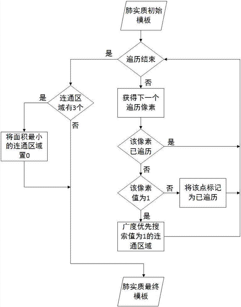
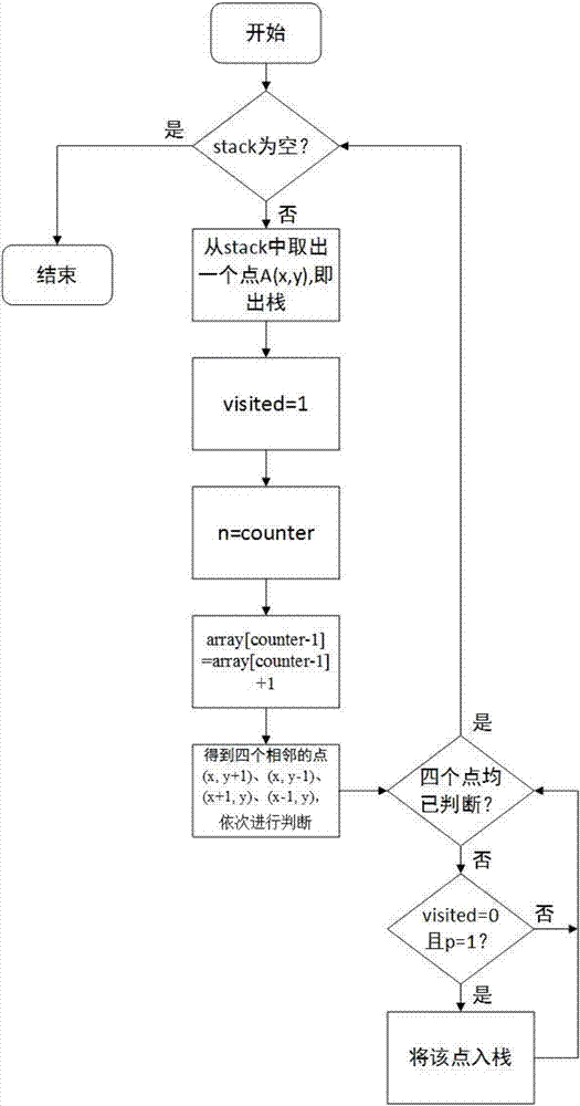
[0063] Embodiment 1, the lung CT image lung parenchyma template tracheal region elimination method based on breadth-first search, such as Figure 1~4 As shown, it includes three steps of traversing the pixels of the lung parenchyma template, searching for connected regions and removing connected regions with the smallest area.

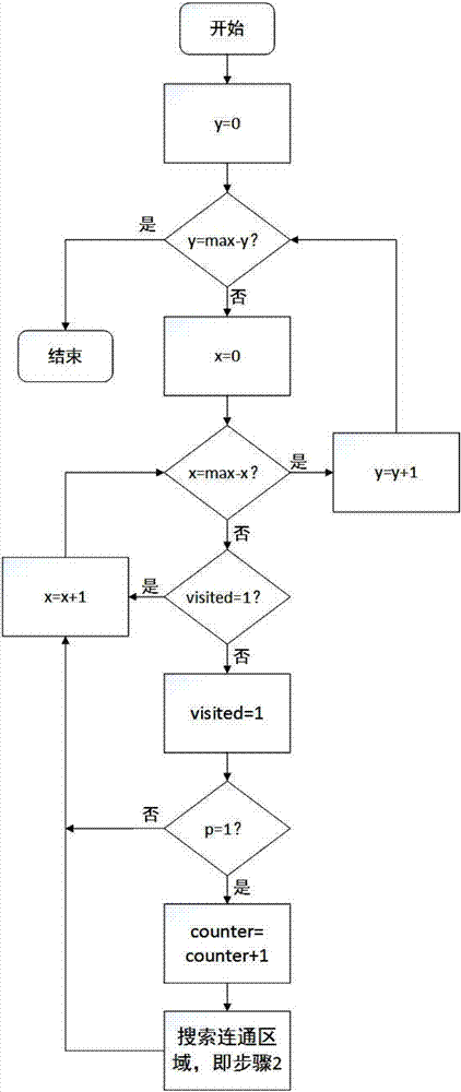
[0064] instruction manual: figure 1 What embodies is the overall process of the present invention, figure 2 embodies the figure 1 The part of traversing pixels in is equivalent to the main loop, including figure 1 In the steps of "detecting the end of traversal", "obtaining the next traversed pixel", "judging that the pixel has been traversed", "judging that the value of the pixel is 1" and "marking the point as traversed".

[0065] Take the pixel point in the lower left corner of the lung parenchyma template as the coordinate origin (0, 0), establish a two-dimensional x-y coordinate system, and each coordinate point represents a pixel, assuming th...


PUM


Abstract

The invention provides a CT image pulmonary parenchyma template weasand elimination method based on breadth-first search. The method comprises the steps that (1) traversal starts from traversal pixels in a pulmonary parenchyma template; (2) whether the traversal is completed or not is determined, and if so, the step (8) is carried out, otherwise the step (3) is carried out; (3) a next traversal pixel is acquired; (4) whether the pixel is traversed is determined, and if so, the step (2) is carried out, otherwise the step (5) is carried out; (5) whether the pixel value is 1 is determined, and if so, the step (6) is carried out, otherwise the step (7) is carried out; (6) in a communication area with the breadth-first search pixel value of 1, the step (2) is carried out; (7) the point is marked as traversed, and then the step (2) is carried out; (8) whether the number of communication areas is three is determined, and if so, the step (9) is carried out, otherwise a final pulmonary parenchyma template is acquired; and (9) the pixel value of all pixels in the smallest communication area is set to 0 to acquire the final pulmonary parenchyma template.

Description

technical field [0001] The invention relates to a method for eliminating the trachea area of ​​a lung parenchyma template in a CT image of a lung based on breadth-first search. Background technique [0002] With the development and maturity of medical imaging technology, medical imaging plays an important role in the diagnosis of lung diseases. CT uses precisely collimated X-ray beams, γ-rays, ultrasound, etc., together with highly sensitive detectors, to perform cross-sectional scanning around a certain part of the human body one by one. It is already an important means of lung disease inspection. In computer-aided detection, it is necessary to perform feature extraction, analysis, and recognition on lung CT images. In addition to lung parenchyma, there are some other organs and tissues in ordinary lung CT images, as well as the background of CT images. The feature extraction of a CT image will affect the accuracy of recognition. Therefore, it is necessary to segment the l...

Claims


Application Information

Patent Timeline
IPC IPC(8): G06T7/11G06T7/187
CPCG06T2207/10081G06T2207/30061
Inventor 金心宇刘俊洋
Owner ZHEJIANG UNIV
Why Eureka
Try Eureka