Nuclear imaging using three-dimensional gamma particle interaction detection

a technology of gamma particle interaction and nuclear imaging, applied in the field of nuclear imaging, to achieve the effect of reducing uncertainty, increasing the resolution of composite images, and ensuring the accuracy of the imag

Inactive Publication Date: 2005-02-03
SIEMENS MEDICAL SOLUTIONS USA INC
View PDF2 Cites 57 Cited by
  • Summary
  • Abstract
  • Description
  • Claims
  • Application Information

AI Technical Summary

Benefits of technology

The present invention significantly reduces the uncertainty associated with each line of interaction generated in PET-type, coincident interaction-based imaging and therefore increases the resolution of the composite image that is generated. The invention accomplishes this by taking advantage of the ability to determine the depth of each individual gamma ray / electron interaction within a semiconductor detector, which in turn allows one to determine the position of the interaction within three-dimensional space.

Problems solved by technology

However, because all gamma ray / electron interactions within each array of semiconductors occur essentially simultaneously (due to the fact that the scattering occurs at the speed of light), it is not possible from this information alone to determine the actual sequence of interactions and hence the location of the first gamma interaction (which is more indicative of the true direction from which the gamma ray originally came than the average, centroid of interaction events).

Method used


Image

  • Nuclear imaging using three-dimensional gamma particle interaction detection
  • Nuclear imaging using three-dimensional gamma particle interaction detection
  • Nuclear imaging using three-dimensional gamma particle interaction detection

Examples


Embodiment Construction

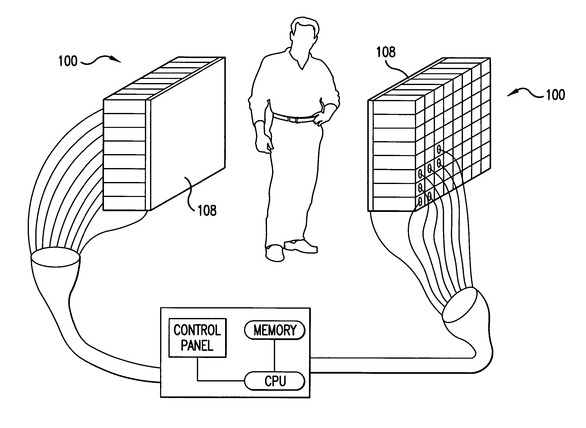
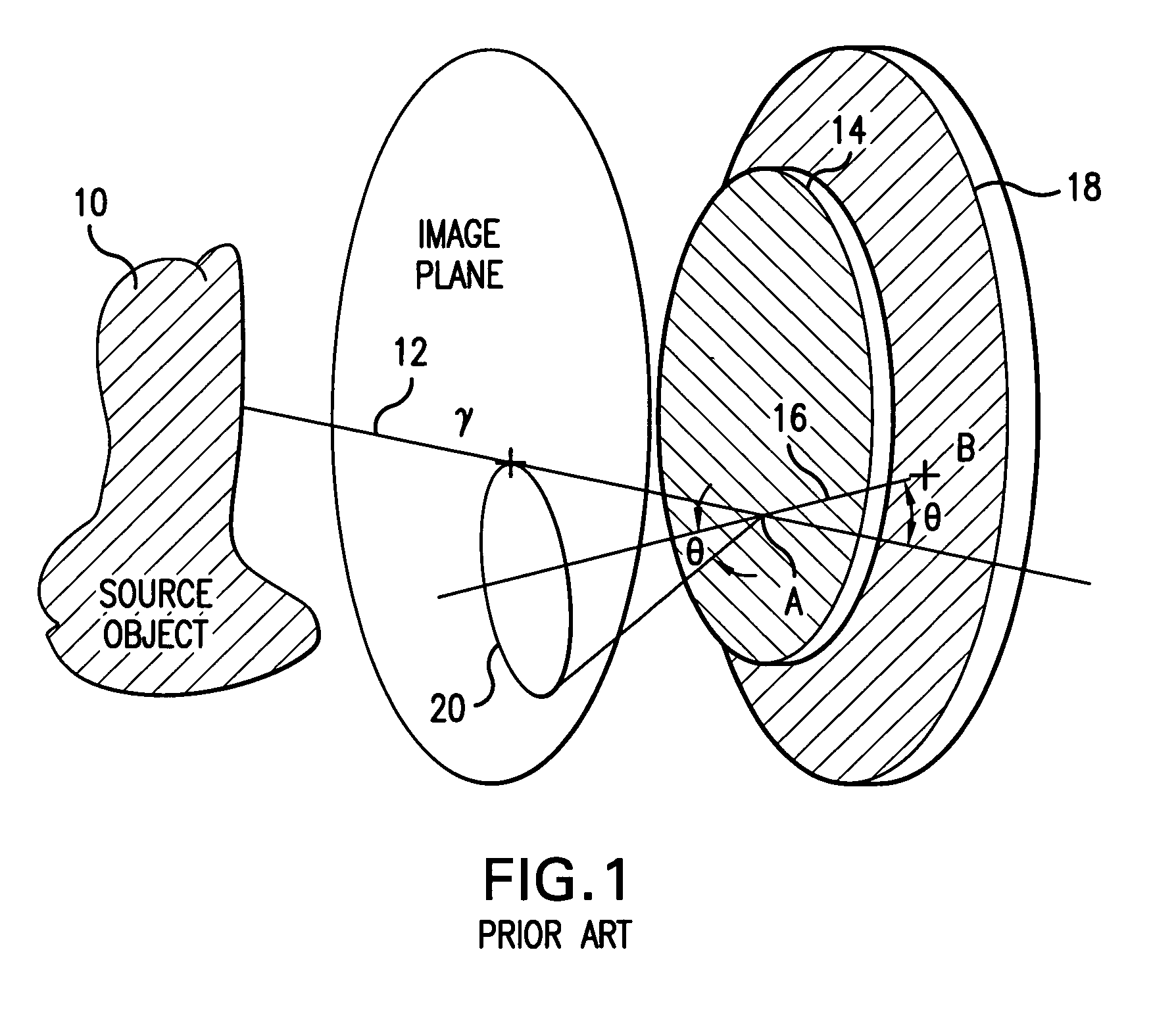
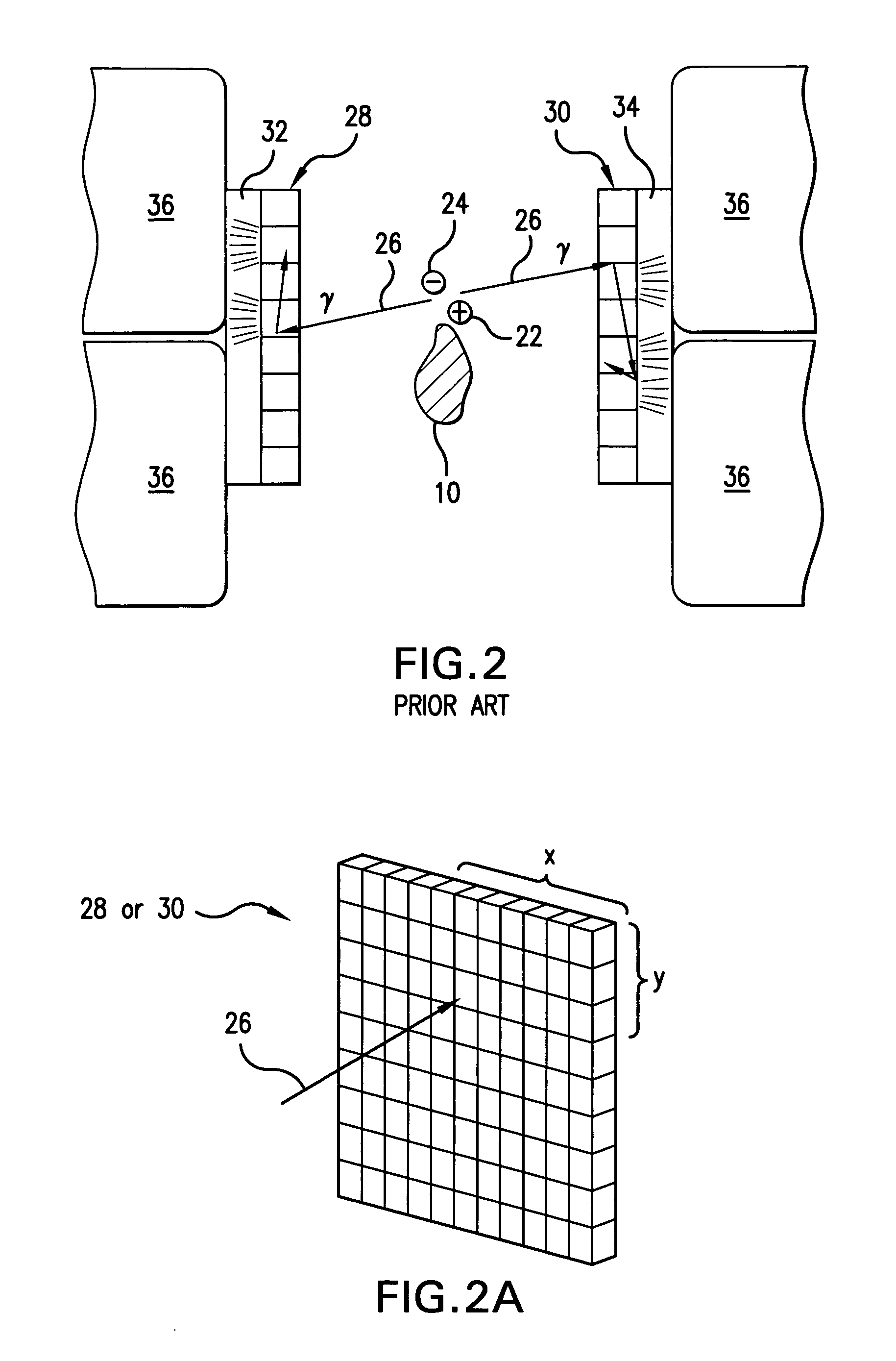
The imaging method and apparatus of the invention utilize a pair of opposing, semiconductor-based gamma interaction detectors that are arranged somewhat similarly to the prior art, scintillation-based detectors shown in FIG. 2. Each detector 100 (FIG. 3) is constructed as an array of individual semiconducting elements or “pixels”102, which preferably are cadmium zinc telluride, CdZnTe, and each detector has on the order of ten by ten such pixels. Each element or pixel 102, which is on the order of one millimeter by one millimeter in height and width and on the order of one centimeter in length, has an electrode at each end thereof. Preferably, the detector is constructed with individual coplanar grid anodes 104 (and their associated leads 106) bonded to the individual ends of the detecting elements forming one surface of the detector 100 and a single continuous cathode 108 bonded across all the opposite ends of the semiconductor detecting elements 102 in a gamma interaction-detecti...


PUM


Abstract

An improved method of, and apparatus for, nuclear imaging takes advantage of the ability to determine the depth of gamma ray / electron interaction within a semiconducting gamma ray detector to determine the (most highly probable) location of the first gamma ray / electron interaction within the detector. Lines of interaction constructed between opposing detector arrays, extending between the location of the first gamma ray / electron interaction in each detector associated with the coincident detection of gamma radiation, permits a positron-emitting object of interest to be imaged according to protocols known in the art, but with better spatial resolution than previously believed to have been known.

Description

FIELD OF THE INVENTION The present invention relates generally to nuclear imaging, e.g., of the sort used in nuclear medicine. More particularly, the invention relates to an improved method of, and apparatus for, imaging using coincident detection of gamma ray interactions, with two different spaced detectors, as typically used in positron emission tomography (PET) scanning. BACKGROUND OF THE INVENTION In general, nuclear imaging that is based on detecting gamma rays is well known. In a Compton scatter camera, for example, basic operating principles of which are illustrated schematically in FIG. 1, a source object of interest 10, e.g., a particular organ within the human body, is caused to be impregnated with a radioisotope which emits gamma radiation. A gamma photon travels along a first path of travel 12 until it interacts with an electron of an atom within the detector at location A within a first detector 14. The gamma photon is then deflected or scattered by a Compton scatter...

Claims


Application Information

Patent Timeline
Patent Type & Authority Applications(United States)
IPC IPC(8): G01T1/24G01T1/29H01L25/00
CPCG01T1/2985
Inventor PERSYK, DENNIS E.ENGDAHL, JOHN C.
Owner SIEMENS MEDICAL SOLUTIONS USA INC
Why Eureka
Try Eureka