Medical image processing method and equipment

A medical image and processing method technology, applied in the field of medical image processing, can solve problems such as low accuracy and poor robustness, and achieve the effects of improving accuracy, reducing artifacts, and improving signal-to-noise ratio.

Active Publication Date: 2017-09-01
SHANGHAI UNITED IMAGING HEALTHCARE
View PDF5 Cites 23 Cited by
  • Summary
  • Abstract
  • Description
  • Claims
  • Application Information

AI Technical Summary

Problems solved by technology

[0009] In view of this, the embodiment of this solution provides a medical image processing method and equi

Method used


Image

  • Medical image processing method and equipment
  • Medical image processing method and equipment
  • Medical image processing method and equipment

Examples


Embodiment 1

[0068] An embodiment of the present invention provides a medical image processing method. The medical image processing method can be implemented through an application program APP, and terminals such as computers and medical workstations can obtain corresponding medical image processing functions by installing the application program.

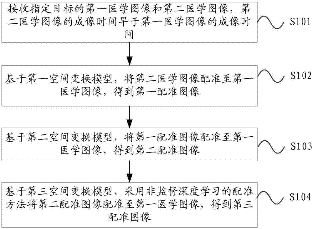
[0069] figure 1 It is an example diagram of the first flow chart of the medical image processing method provided by the embodiment of the present invention. Such as figure 1 As shown, in this embodiment, the medical image processing method may include the following steps:

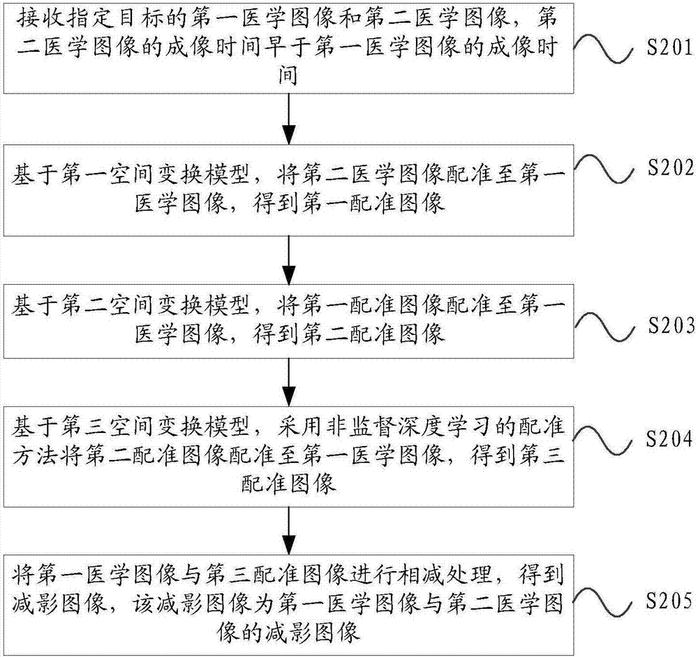
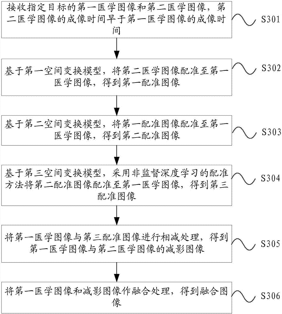
[0070] S101. Receive a first medical image and a second medical image of a specified target, where the imaging time of the second medical image is earlier than the imaging time of the first medical image.

[0071] Optionally, the interval between the imaging time of the first medical image and the imaging time of the second medical image is generally not less than 24 hour...

Embodiment 2

[0148] An embodiment of the present invention provides a medical image processing device, which can implement the steps of the medical image processing method in the foregoing embodiments.

[0149] Figure 5 The functional block diagram of the medical image processing device provided by the embodiment of the present invention. Such as Figure 5 As shown, in this embodiment, the medical image processing device includes:

[0150] A receiving module 510, configured to receive a first medical image and a second medical image of a specified target, the imaging time of the second medical image is earlier than the imaging time of the first medical image;

[0151] The first registration module 520 is configured to register the second medical image to the first medical image based on the first spatial transformation model to obtain a first registration image;

[0152] The second registration module 530 is configured to register the first registration image to the first medical image...

Embodiment 3

[0163] An embodiment of the present invention provides a medical image processing device, which includes: a processor; a memory for storing instructions executable by the processor; the processor is configured to: receive a first medical image and a second medical image of a specified target, The imaging time of the second medical image is earlier than the imaging time of the first medical image; based on the first spatial transformation model, the second medical image is registered to the first medical image to obtain the first registration image; based on the second spatial transformation model , register the first registration image to the first medical image to obtain the second registration image; based on the third spatial transformation model, use the registration method of unsupervised deep learning to register the second registration image to the first medical image image to obtain a third registered image.

[0164] Wherein, the medical image processing device may be ...


PUM


Abstract

The invention provides a medical image processing method and equipment. According to the method, a first medical image and a second medical image of a designated target are received; first registration, second registration and registration based on unsupervised depth learning for the second medical image are carried out sequentially to acquire a third registration image, alignment of main structures of the two images and alignment of areas of interests of the lung can be realized through first registration and second registration, based on registration based on unsupervised depth learning and characteristics of the lung containing multiple blood vessels, fine blood vessel structures of the lung are extracted, and precise registration based on unsupervised depth learning is carried out for the fine blood vessel structures. The method is advantaged in that medical image registration precision is improved, more strong robustness is realized, and problems of low precision and poor robustness existing in medical image registration in the prior art are solved.

Description

[0001] 【Technical field】 [0002] The solution relates to the field of medical image processing, in particular to a medical image processing method and equipment. [0003] 【Background technique】 [0004] Medical imaging technology has experienced a development process from static to dynamic, from form to function, from plane to three-dimensional, especially driven by imaging hardware, the development of medical imaging technology provides clinical medicine with images of various forms and functions information. At present, the research objects of medical image analysis are becoming more and more extensive. Automatic, accurate and quantitative computer-aided image analysis can be used to help clinicians and researchers in colleges and universities to accurately process massive image information. [0005] In medical image analysis, it is often necessary to analyze several images of the same patient together, so as to obtain comprehensive information of the patient and improve th...

Claims


Application Information

Patent Timeline
IPC IPC(8): G06T7/33
CPCG06T7/33G06T2207/10081G06T2207/20081G06T2207/20221G06T2207/30061G06T2207/30101
Inventor 严计超李强
Owner SHANGHAI UNITED IMAGING HEALTHCARE
Why Eureka
Try Eureka