Wireless capsule endoscope image display method for highlighting vessel mutation and projection

A technology of capsule endoscope and display method, which is applied in the directions of image analysis, image data processing, graphic image conversion, etc., and can solve problems such as low efficiency, aggravated disease, and time-consuming

Inactive Publication Date: 2017-09-05
CHONGQING JINSHAN MEDICAL INSTR CO LTD
View PDF6 Cites 2 Cited by
  • Summary
  • Abstract
  • Description
  • Claims
  • Application Information

AI Technical Summary

Problems solved by technology

And these massive image data must be manually interpreted by professional medical personnel, which is labor-intensive, inefficient, and time-consuming
Moreover, it is also possible that the lesion area is not so obvious in the pictures of the lesion area found in many images, which increases the misjudgment of the patient's condition, not only aggravates the condition, but also delays the treatment.

Method used


Image

  • Wireless capsule endoscope image display method for highlighting vessel mutation and projection
  • Wireless capsule endoscope image display method for highlighting vessel mutation and projection
  • Wireless capsule endoscope image display method for highlighting vessel mutation and projection

Examples


Embodiment Construction

[0035] Embodiments of the present invention are described in detail below, examples of which are shown in the drawings, wherein the same or similar reference numerals designate the same or similar elements or elements having the same or similar functions throughout. The embodiments described below by referring to the figures are exemplary only for explaining the present invention and should not be construed as limiting the present invention.

[0036] In describing the present invention, it should be understood that the terms "longitudinal", "transverse", "upper", "lower", "front", "rear", "left", "right", "vertical", The orientation or positional relationship indicated by "horizontal", "top", "bottom", "inner", "outer", etc. are based on the orientation or positional relationship shown in the drawings, and are only for the convenience of describing the present invention and simplifying the description, rather than Nothing indicating or implying that a referenced device or elem...


PUM


Abstract

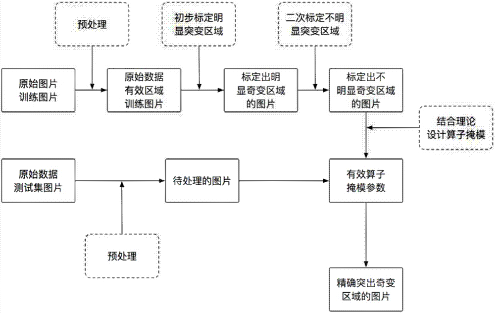
The invention discloses a display method for highlighting a vessel mutation and a projection. The wireless capsule endoscope image display method comprises steps of 1) obtaining original image data of an intestine and a stomach which is obtained by a wireless capsule endoscope, wherein the a doctor determines that blood mutation or the projection is not obvious according to the original image data of the stomach and intestine, 2) finding out a singular point which is not obvious in mutation, designing an operator mask layer for enhancing vessel mutation according to theories of image rim enhancement and image texture enhancement, and 3) using modified various parameters to perform corresponding processing on images of various parts to obtain an image highlighting the mutation part for doctors to detect according to images of various parts. The wireless capsule endoscope image display method can perform processing of highlighting the vessel mutation on the wireless endoscope image, enables the doctor to better diagnose patients and is applicable to computer medical image processing.

Description

technical field [0001] The invention relates to the field of computer image processing, in particular to a display method for highlighting vascular anomalies and protruding parts in wireless capsule endoscope images in the medical field. Background technique [0002] As an important new technology, wireless capsule endoscopy images are widely used in the diagnosis of human gastrointestinal diseases. It can reach any position in the digestive tract, overcomes the shortcomings of traditional endoscopy that cannot detect small intestinal diseases, and is non-invasive and well tolerated. Although the technology has been continuously improved with the advancement of science and technology, it still faces some difficulties in practical application. The capsule endoscope stays in the digestive tract for an average of 8 hours. During this period, its camera system will automatically capture the entire digestive tract scene, and can collect a total of 50,000 to 60,000 color digestiv...

Claims


Application Information

Patent Timeline
Patent Type & Authority Applications(China)
IPC IPC(8): G06T7/00G06T3/00
CPCG06T7/0012G06T2207/10024G06T2207/10068G06T2207/20104G06T2207/30092G06T3/04
Inventor 张寻汪成亮郭凌白家莲严莎莎
Owner CHONGQING JINSHAN MEDICAL INSTR CO LTD
Why Eureka
Try Eureka