Generating contrast-enhanced image data based on multi-energy x-ray imaging

An image data, X-ray technology, applied in the field of X-ray imaging, can solve problems such as reducing the application range

Inactive Publication Date: 2017-09-05
SIEMENS HEALTHCARE GMBH
View PDF6 Cites 6 Cited by
  • Summary
  • Abstract
  • Description
  • Claims
  • Application Information

AI Technical Summary

Problems solved by technology

The scope of application of the imaging methods described above is significantly reduced due to the specific dose constraints that must be observed

Method used


Image

  • Generating contrast-enhanced image data based on multi-energy x-ray imaging
  • Generating contrast-enhanced image data based on multi-energy x-ray imaging
  • Generating contrast-enhanced image data based on multi-energy x-ray imaging

Examples


Embodiment Construction

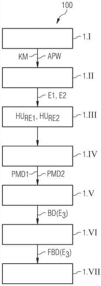
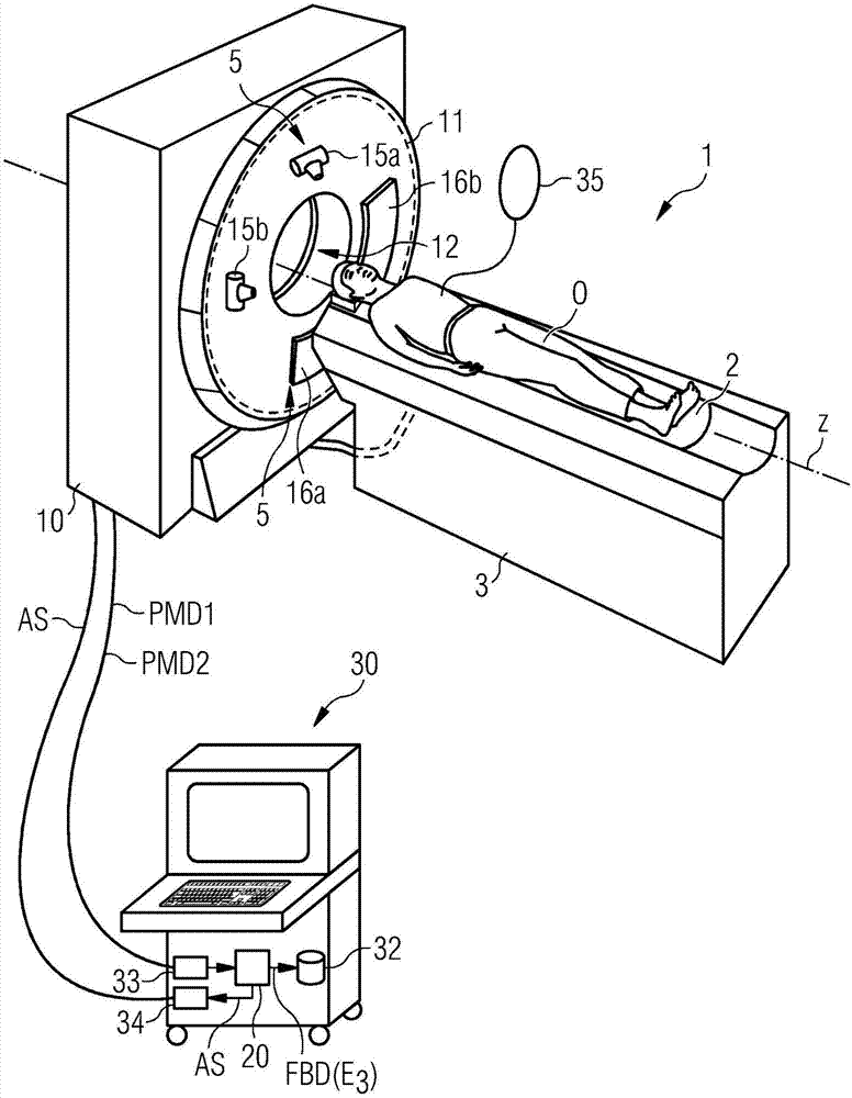
[0046] figure 1 A flowchart 100 is shown, which shows a dynamic CT imaging method by means of the so-called dual-energy technique according to an exemplary embodiment of the invention, in which contrast-enhanced image data is generated from obese patients. In the dynamic imaging method by means of the dual-energy technique, a dynamic time-resolved acquisition of two projection measurement datasets PMD1, PMD2 is performed, each with a different mean energy E 1 ,E 2 by having a different X-ray energy spectrum R E1 , R E2 generated by X-ray radiation. In order to use a different X-ray energy spectrum R E1 , R E2 X-ray radiation is generated, for example two X-ray sources 15a, 15b can be used (see image 3 ), which at different X-ray energies E 1 ,E 2 or X-ray spectroscopy R E1 , R E2 emit X-ray radiation.

[0047] In the context of a dynamic imaging method, in step 1.1 first data relating to the dimensions of the object to be examined (eg a patient) is acquired. This ...


PUM


Abstract

An X-ray imaging method (100) is for generating contrast-enhanced image data (BD) relating to an examination region (FOV) of an object (O) to be examined. Perferably a CT-X ray imaging method is disclosed. In an embodiment of the method, first contrast-agent influenced measured X-ray projection data (PMD1) with a first X-ray energy spectrum (RE1) and at least one set of second contrast-agent influenced measured X-ray projection data (PMD2) with a second X-ray energy spectrum (RE2) are acquired from the examination region. Subsequently, image data assigned to a third X-ray energy spectrum with a third mean energy (E3), based on the first and at least second measured X-ray projection data is reconstructed based on the first and at least second measured X-ray projection data that has been acquired. A mean energy of the first X-ray energy spectrum and a mean energy of the second X-ray energy spectrum are selected as a function of a dimension parameter value of the object that is to be examined. The invention also describes an image data generating device (20) and a computer tomography system (1).

Description

technical field [0001] The invention relates to an X-ray imaging method for generating contrast-enhanced image data. The invention also relates to an image data generating device. Furthermore, the invention relates to a computer tomography system. Background technique [0002] With the aid of modern imaging methods, two-dimensional or three-dimensional image data are frequently generated which can be used to visualize the imaged examination object and which can also be used for other applications. [0003] Imaging methods are generally based on the detection of X-ray radiation, where "projection measurement data" are generated. For example, projection measurement data can be acquired by means of a computer tomography system (CT system). In a CT system, the combination of an X-ray source and an X-ray detector arranged opposite said source and arranged on a gantry usually rotates around a measurement space, examining an object (without loss of generality, hereinafter mostly...

Claims


Application Information

Patent Timeline
IPC IPC(8): G06T11/00G06T5/00A61B6/00A61B6/03
CPCA61B6/00A61B6/032A61B6/42A61B6/5205A61B6/5258A61B6/54G06T11/00G06T11/005G06T2207/10081G06T2207/10116G06T5/70A61B6/4014A61B6/405A61B6/504A61B6/507A61B6/4241A61B6/482A61B6/4007A61B6/481
Inventor T·弗洛尔B·施密特
Owner SIEMENS HEALTHCARE GMBH
Why Eureka
Try Eureka