Radiomics-based brain glioma grading prediction method

A technology of glioma and radiomics, applied in image analysis, image enhancement, image data processing, etc., can solve the problem of uncertain metastasis location, unfavorable treatment plan and grading prediction of glioma grade, glioma Unclear organizational boundaries and other issues to achieve the effect of alleviating pain and reducing errors

Inactive Publication Date: 2017-09-29
ZHENGZHOU UNIV
View PDF0 Cites 23 Cited by
  • Summary
  • Abstract
  • Description
  • Claims
  • Application Information

AI Technical Summary

Problems solved by technology

[0002] The technology based on medical imaging has been applied to the diagnosis and staging of cancer. Due to the characteristics of continuous angiogenesis, tissue invasion and metastasis of glioma, the boundary between normal tissue and glioma tissue is not clear, and the location of metastasis is uncertain. Determining the factors leading to the prognosis prediction of glioma becomes a severe challenge in clinical research
At present,

Method used


Image

  • Radiomics-based brain glioma grading prediction method
  • Radiomics-based brain glioma grading prediction method
  • Radiomics-based brain glioma grading prediction method

Examples


Embodiment Construction

[0021] The technical solutions of the present invention will be described in further detail below through specific implementation methods.

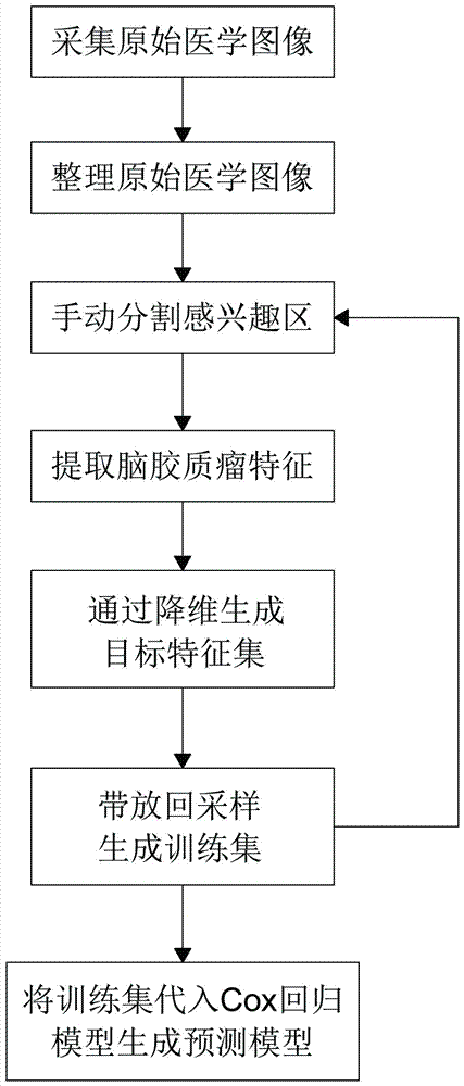
[0022] Such as figure 1 As shown, a radiomics-based glioma grade prediction method includes the following steps:

[0023] Step 1: Acquire the original medical image of glioma by conventional magnetic resonance imaging method, and unify the image format and size of the collected original medical image and deprivate the data to obtain the medical image sample of glioma Group;

[0024] Wherein, the conventional magnetic resonance imaging method includes one or more combinations of T1-weighted imaging, T2-weighted imaging, fluid-attenuated inversion recovery (FLAIR) imaging or enhanced T1-weighted imaging;

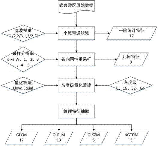
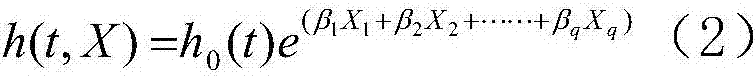
[0025] Step 2, select one of the glioma medical image samples, and manually segment the ROI in the glioma medical image sample; specifically, the ROI segmentation results must be confirmed by two radiologists. If they are different, tw...


PUM


Abstract

The present invention provides a method for grading and predicting brain gliomas based on radiomics, comprising the following steps: step 1, collecting original medical images of brain gliomas by conventional magnetic resonance imaging methods, and performing image processing on the collected original medical images Format and size unification and data de-privacy processing, to obtain a glioma medical image sample group; step 2, select one of the glioma medical image samples, and manually segment the sensory information in the glioma medical image sample region of interest; step 3, extracting the features of glioma in the region of interest; step 4, performing dimensionality reduction on the features of glioma extracted in step 3, selecting effective features, and generating a target feature set; step 5, in the brain Sampling with replacement is performed in the glioma medical image sample group, and steps 2 to 4 are repeated to generate a training set; step 6, the prediction model is trained using the Cox regression model according to the training set to generate a prediction model.

Description

technical field [0001] The present invention relates to radiomics auxiliary diagnosis technology, in particular, relates to a radiomics-based glioma grade prediction method. Background technique [0002] The technology based on medical imaging has been applied to the diagnosis and staging of cancer. Due to the characteristics of continuous angiogenesis, tissue invasion and metastasis of glioma, the boundary between normal tissue and glioma tissue is not clear, and the location of metastasis is uncertain. Determining the factors leading to the prediction of the prognosis of glioma has become a severe challenge in clinical research. At present, in the clinical aspect of glioma imaging, radiologists mainly make subjective and qualitative judgments on the grading diagnosis of gliomas in the examination results based on personal knowledge and experience, and the results of the judgments also contain simple opinions. Quantitative information cannot fully describe tumor informatio...

Claims


Application Information

Patent Timeline
IPC IPC(8): G06T7/00G06F19/00
CPCG06T7/0012G06T2207/10088G06T2207/20081G06T2207/30016G06T2207/30096
Inventor 林予松刘博吴亚平庞海波赵哲王梅云
Owner ZHENGZHOU UNIV
Why Eureka
Try Eureka