A placenta potential cell and a culture method thereof
A technology of potential cells and culture methods, which is applied in the field of potential cells derived from animal placenta and its culture, and can solve the problems of poor growth and proliferation of cells
- Summary
- Abstract
- Description
- Claims
- Application Information
AI Technical Summary
Problems solved by technology
Method used
Image
Examples
Embodiment 1
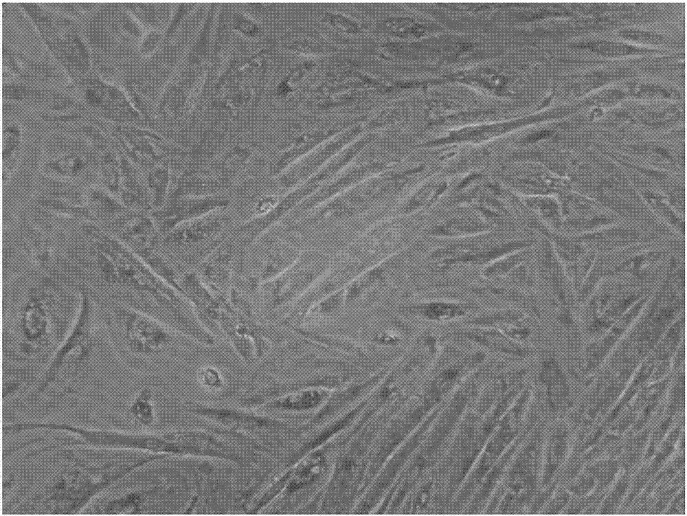
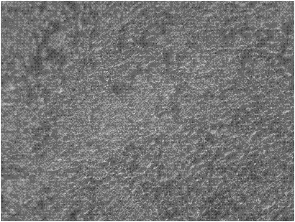
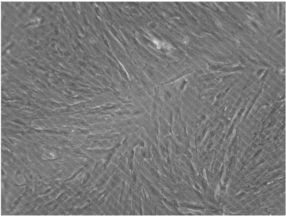
[0110] Embodiment 1 Culture human placenta cell in vitro
[0111] To obtain human placenta, first put the placental tissue into 4°C pre-cooled phosphate-buffered saline (PBS) containing double antibodies (penicillin and streptomycin), wash 3 times, and then cut the large tissue block into 2mm For small tissue pieces, wash twice with cold PBS containing double antibodies, and put the washed tissue pieces into 0.25% neutral protease solution or 1% collagenase solution prepared with sterile PBS for digestion, and the condition is constant temperature at 37°C Oscillate for 1 hour, and then carry out experiments according to the following methods.
[0112] Pipette the tissue block repeatedly with a pipette or a pipette, or pour the tissue block together with the digestive solution onto a stainless steel filter, and grind the tissue block with a syringe plunger until the tissue block is completely dispersed and becomes a single cell.
[0113] This contains cells and a digestive enz...
Embodiment 2
[0124] Example 2 Formation of Induced Differentiation of Adipose Tissue, Bone and Cartilage
[0125] see image 3 , continue to culture the placental cells of the experimental group, adding three inducers that form bone, cartilage, and fat. After two weeks, adipose tissue, bone, and cartilage are formed.
[0126] see Figure 4-5 , using different staining methods, under the microscope, to form bone, cartilage, and adipose tissue.
Embodiment 3
[0127] Example 3, identification of cell surface markers
[0128] Referring to Fig. 7, the same method as in Example 1 was used for culturing, and a control group and an experimental group were set up, and no substance of the present invention was added to the control group. After 30 days of culture, the cell surface markers were measured by flow cytometry. see Figures 7A-7I , the experimental results are shown below. Figure 7A Show CD90>95%, Figure 7B Show CD34 Figure 7C show CD14 Figure 7D show CD45 Figure 7E show CD19 Figure 7F Show CD105>95%, Figure 7G Show CD31 Figure 7H Show CD73>95%, Figure 7I Show HLA-DR<2%. The cells in the experimental group express CD90, CD73, and CD105, but do not express CD45, CD14, and HLA-DR, and the control group does not express them.
PUM
 Abstract
Description
Claims
Application Information
 - R&D
- Intellectual Property
- Life Sciences
- Materials
- Tech Scout
- Unparalleled Data Quality
- Higher Quality Content
- 60% Fewer Hallucinations
Browse by: Latest US Patents, China's latest patents, Technical Efficacy Thesaurus, Application Domain, Technology Topic, Popular Technical Reports.
© 2025 PatSnap. All rights reserved.Legal|Privacy policy|Modern Slavery Act Transparency Statement|Sitemap|About US| Contact US: help@patsnap.com


