Method for establishing standard median sagittal plane by applying bone markers for craniofacial deformity
A sagittal and craniomaxillofacial technology, applied in the field of maxillofacial plastic surgery, can solve the problems of different types of deformities, no unity, and large errors in the median sagittal plane, so as to improve accuracy and reduce operation time.
- Summary
- Abstract
- Description
- Claims
- Application Information
AI Technical Summary
Problems solved by technology
Method used
Image
Examples
Embodiment 1
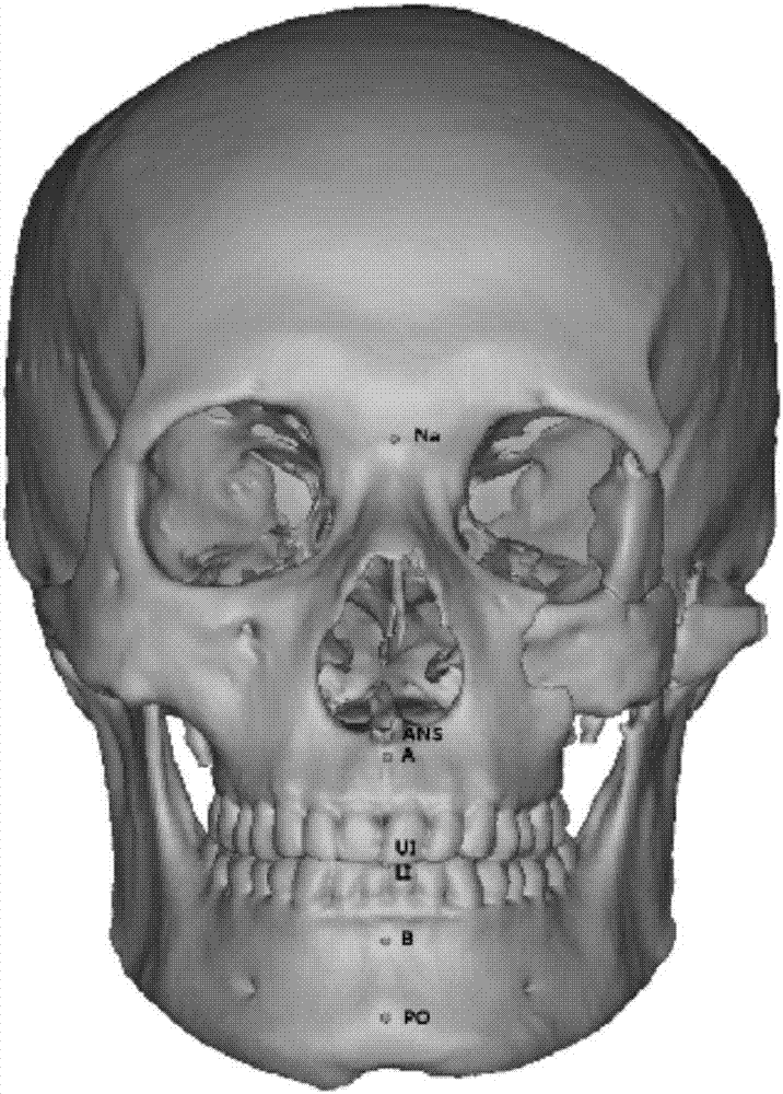
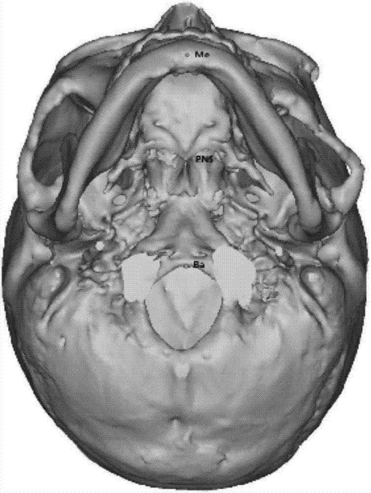
[0030] The present invention proposes a new method for determining the median sagittal plane by applying bony landmarks according to different types of cranio-maxillofacial deformities, and applying data image processing software (Mimics software, geomagic software) to establish and apply bony landmarks to locate the median sagittal plane The specific method is as follows:
[0031] (1) Data acquisition: Spiral CT data of 10 patients with cranio-maxillofacial deformities (cranio-maxillofacial fractures, tumors, bone defects) in West China Stomatological Hospital in 2015 were selected.
[0032] A 16-slice thin-layer spiral CT (Philips MX16 EVO CT, Holland) was used, and the parameters were set to: voltage 120kv, current 7700mAs, pixel size: 512×512, layer spacing: 0.5mm, field of view: 250mm, layer thickness: 1.0mm). During the shooting, the patient also asked for a relaxed expression, with both eyes looking forward, and the occlusion in the mouth was cusp interlacing, without chewin...
Embodiment 2
[0062] Patient Zhang, male, 19 years old. High falls in January. Diagnosis: Comminuted fracture of mandibular chin; fracture of left condyle.
[0063] The patient only had a mandibular fracture, which was characterized by the displacement of the mandibular landmarks along with the fracture, and the remaining landmarks remained unchanged.
[0064] Determine the mid-sagittal plane: Generally speaking, the base of the nose is selected for the upper area, the anterior nasal spine or the upper central incisor point is preferred for the lower area, and the skull base or posterior nasal spine is preferred for the posterior area. If the anterior nasal spine or the upper central incisor point is obviously deviated, consider using a posterior nasal spine instead.
[0065] After determining the median sagittal plane, locate the anterior chin, chin or incisor point, and guide the fracture block so that these three landmark points are located on the median sagittal plane. Specific steps are as...
Embodiment 3
[0072] The patient Jiang Mou, male, 43 years old. Injured for 2 days. Diagnosis: Left zygomatic orbital maxilla and zygomatic arch fracture.
[0073] The patient had a unilateral middle fracture, which was characterized by no deviation of all landmarks.
[0074] Determine the mid-sagittal plane: choose the nasal root point in the face area, the anterior chin point or the chin point in the subface area, and the skull base point in the posterior area. By determining the median sagittal plane to mirror the normal side to the fractured side, the reduction of the fractured block on the fractured side can be known. Specific steps are as follows:
[0075] (1) Spiral CT was taken in the supine position of the patient, and the data was imported into MIMICS software to reconstruct the skull image and segment the fracture.
[0076] (2) The patient had a unilateral middle fracture, and the occlusal relationship was basically normal. Choose the nasal root point, the anterior chin point, and the...
PUM
 Abstract
Description
Claims
Application Information
 - R&D Engineer
- R&D Manager
- IP Professional
- Industry Leading Data Capabilities
- Powerful AI technology
- Patent DNA Extraction
Browse by: Latest US Patents, China's latest patents, Technical Efficacy Thesaurus, Application Domain, Technology Topic, Popular Technical Reports.
© 2024 PatSnap. All rights reserved.Legal|Privacy policy|Modern Slavery Act Transparency Statement|Sitemap|About US| Contact US: help@patsnap.com


