Method for extracting cardiovascular vessels from CTA images, device, apparatus and storage medium

A cardiovascular and image-based technology, which is applied in the fields of equipment and storage media, methods for extracting cardiovascular systems, and devices, can solve problems such as the structure of pulmonary vessels and the inability to extract cardiovascular methods, and achieve the effect of improving the level and ability of solving

Active Publication Date: 2017-12-08
SHENZHEN INST OF ADVANCED TECH
View PDF5 Cites 9 Cited by
  • Summary
  • Abstract
  • Description
  • Claims
  • Application Information

AI Technical Summary

Problems solved by technology

[0003] The object of the present invention is to provide a method, device, equipment and storage medium for extracting cardiovascular in CTA images, aiming to solve the problem of the presence of lungs due to the inability of the prior art to provide an effective method for extracting cardiovascular in CTA images. blood vessel problems

Method used


Image

  • Method for extracting cardiovascular vessels from CTA images, device, apparatus and storage medium
  • Method for extracting cardiovascular vessels from CTA images, device, apparatus and storage medium
  • Method for extracting cardiovascular vessels from CTA images, device, apparatus and storage medium

Examples


Embodiment 1

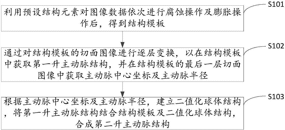
[0023] figure 1 It shows the implementation process of the method for extracting cardiovascular in CTA images provided by Embodiment 1 of the present invention. For the convenience of description, only the parts related to the embodiment of the present invention are shown, and the details are as follows:

[0024] In step S101, the image data is sequentially corroded and dilated by using preset structural elements to obtain a structural template.

[0025] In the embodiment of the present invention, the above-mentioned image data is a coronary angiography image after down-sampling processing. For the large-scale original CTA data, in order to quickly extract the large-scale ascending aorta structure without affecting the accuracy of the structure extraction, it can be Downsample the image size to half its original size. Suppress or weaken some noise in the above image data and some structures that are not related to the aorta, use the preset structural elements, first perform t...

Embodiment 2

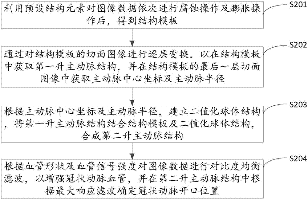
[0038] figure 2 It shows the implementation process of the method for extracting cardiovascular in CTA images provided by Embodiment 2 of the present invention. For the convenience of description, only the parts related to the embodiment of the present invention are shown, and the details are as follows:

[0039] In step S201, the image data is sequentially corroded and expanded by using preset structural elements to obtain a structural template.

[0040] In step S202, the first ascending aorta structure is obtained in the structural template by performing layer-by-layer transformation on the slice image of the structural template, and the coordinates of the center of the aorta and the aortic radius are obtained from the last slice image of the structural template .

[0041] In step S203, a binary sphere structure is established according to the coordinates of the center of the aorta and the radius of the aorta, and the second ascending aorta structure is synthesized by comb...

Embodiment 3

[0050] Figure 4 It shows a schematic structural diagram of extracting a cardiovascular device in a CTA image provided by Embodiment 3 of the present invention. For convenience of description, only the parts related to the embodiment of the present invention are shown. Extracting a cardiovascular device in a CTA image includes:

[0051] The structural template acquisition unit 41 is used to use preset structural elements to sequentially corrode and expand the image data to obtain a structural template. The image data is a coronary angiography image after down-sampling processing, and the structural template is to exclude the lung region Structure.

[0052] In the embodiment of the present invention, the above-mentioned image data is a coronary angiography image after down-sampling processing. For the large-scale original CTA data, in order to quickly extract the large-scale ascending aorta structure without affecting the accuracy of the structure extraction, it can be Downsam...


PUM


Abstract

The invention belongs to the medical image processing technical field and provides a method for extracting cardiovascular vessels from CTA (computed tomography angiography) images, a device for extracting cardiovascular vessels from CTA images, an apparatus for extracting cardiovascular vessels from CTA images and a storage medium. The method includes the following steps that: etching operation and expansion operation are sequentially performed on image data by using preset structural elements, so that a structural template is obtained; layer-by-layer transformation is performed on the sectional images of the structural template, so that a first ascending aorta structure in the structural template can be obtained, aorta center coordinates and an aorta radius are obtained from the last sectional image layer of the structural template; and a binary sphere structure is built according to the aorta center coordinates and the aorta radius, and the first ascending aorta structure is combined with the structural template and the binary sphere structure, so that a second ascending aorta structure can be obtained through combination. With the method, device, apparatus and storage medium of the invention adopted, ascending aortas and the shapes of aortic sinuses at the roots of the ascending aortas can be extracted, and therefore, the structures and shapes of the aortas can be accurately visualized, and the solving level and capability of medical image research on clinical problems can be improved.

Description

technical field [0001] The invention belongs to the technical field of medical image processing, and in particular relates to a method, device, equipment and storage medium for extracting cardiovascular from CTA images. Background technique [0002] Cardiovascular extraction, as a means of image preprocessing, is of great significance for the visualization of clinical diagnosis, interventional surgery planning and interventional treatment process. It is to extract the ascending segment of cardiac aorta and enhance coronary vessels from three-dimensional medical angiography images. Since the CT (Computed Tomography, computerized tomography) coronary angiography (CTA) image of the heart presents complex tissue structure and background noise, the contrast of blood vessels in the heart is very small, and the contrast of the external pulmonary artery is extremely high, which increases the risk from cardiac CTA. The difficulty in extracting the aorta and coronary vessels in the im...

Claims


Application Information

Patent Timeline
Patent Type & Authority Applications(China)
IPC IPC(8): G06T7/00
CPCG06T7/0012G06T2207/30101G06T2207/10081G06T2207/20036G06T7/11G06T7/155G06T2207/20061G06T2207/30048G06T2207/20212G06T7/50G06V2201/03
Inventor 周寿军张保昌李宝林王澄陆培
Owner SHENZHEN INST OF ADVANCED TECH
Why Eureka
Try Eureka