
Preparation method of cancer cell ball and application

A technology of tumor cells and spheres, applied in the field of biomedicine, can solve the problems of high price and easy error of Transwell chamber, and achieve the effect of low cost, fast speed and low cost

Active Publication Date: 2017-12-29
THE FIRST AFFILIATED HOSPITAL OF THIRD MILITARY MEDICAL UNIVERSITY OF PLA
View PDF9 Cites 3 Cited by
  • Summary
  • Abstract
  • Description
  • Claims
  • Application Information

AI Technical Summary

Problems solved by technology

This method is prone to errors in the process of removing cells, and the price of Transwell chambers is generally higher

Method used


Image

  • Preparation method of cancer cell ball and application
  • Preparation method of cancer cell ball and application
  • Preparation method of cancer cell ball and application

Examples


Embodiment 1

[0074] 1. Hanging drop of MHCC97H and U87 into spheres

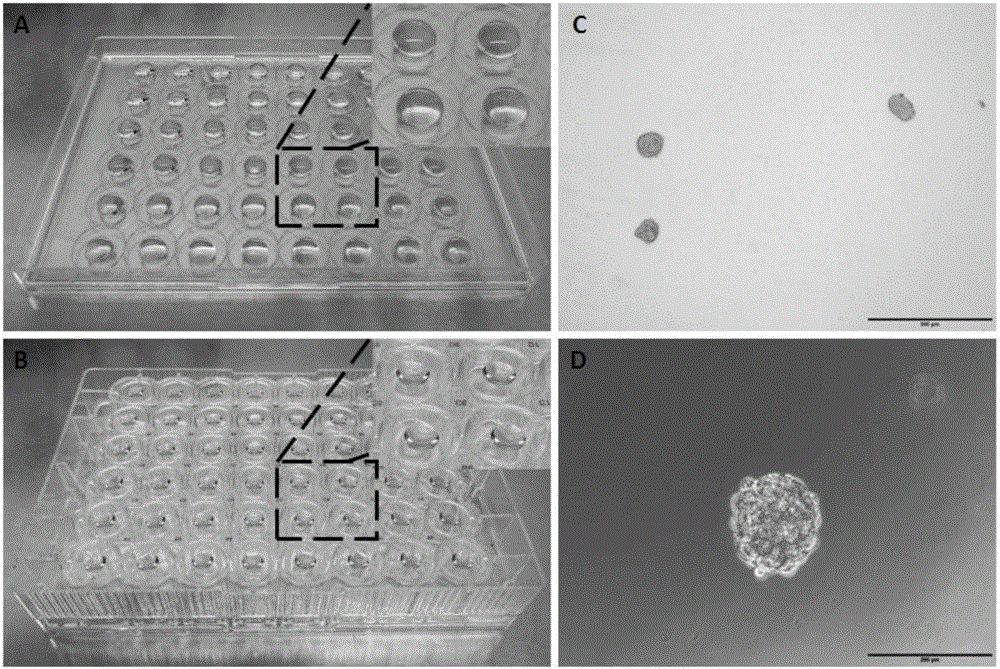
[0075] Resuspend the digested liver cancer cells MHCC97H and glioma cells U87 in DMEM medium, adjust the concentration to 5x 10 3 cells / ml, pipette 20 μl and drop it into a 48-well cell culture plate, one drop per well; then drop the PBS solution drop by drop (50 μl per drop) into the center of the corresponding space on the plate cover, one drop per well; then flip the culture plate on the plate cover above, such as figure 1 shown. 37°C, 5% CO 2 After culturing for 48 hours, spheroidized tumor cells were obtained for the next experiment.

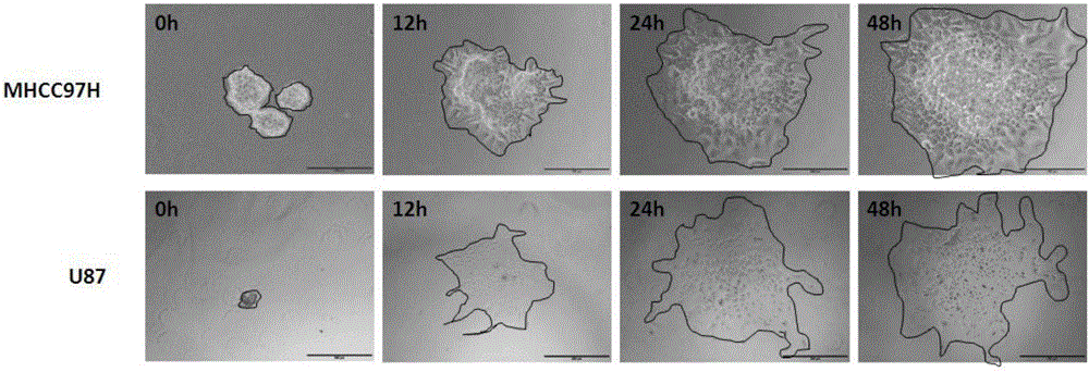
[0076] 2. Matrigel coating of MHCC97H and U87 cell spheres

[0077] Discard the PBS on the cover of the above-mentioned culture plate, place the culture plate upright and put it on ice, add Matrigel pre-thawed on ice one by one, and add 5 μl to each well. Be gentle when adding so as not to damage the cell spheres. Then incubate at 37°C for 30 minutes to make Matrigel coat MHCC97...

Embodiment 2

[0081] 1. MHCC97H hanging drop into ball

[0082] With embodiment 1.

[0083] 2. Matrigel coating of MHCC97H cell spheres

[0084] With embodiment 1.

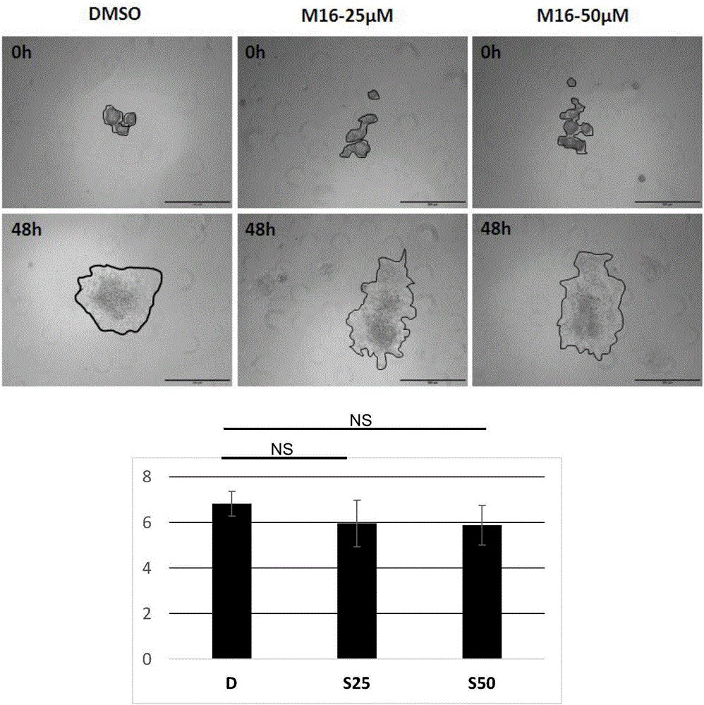
[0085] 3. Add small molecule drug M16

[0086] In the MHCC97H culture plate, add 100 μl of DMEM medium and small molecule drug M16 at concentrations of 25 μM and 50 μM to each well, and incubate for 48 hours. Observed under the microscope, the results are as follows image 3 shown. The difference between the D value and the S value is not large, indicating that M16 does not affect the invasion and migration of MHCC97H cells.

Embodiment 3

[0088] 1. MHCC97H hanging drop into ball

[0089] With embodiment 1.

[0090] 2. Matrigel coating of MHCC97H cell spheres

[0091] With embodiment 1.

[0092] 3. Add small molecule drug M23

[0093] In the MHCC97H culture plate, 100 μl of DMEM medium and the small molecule drug M23 at concentrations of 25 μM and 50 μM were added to each well, and cultured for 48 hours. Observed under the microscope, the results are as follows Figure 4 shown. D value was significantly higher than S value, indicating that M23 can inhibit the invasion and migration of MHCC97H cells.


PUM


Abstract

The invention belongs to the field of a biological medicine, and particularly relates to a preparation method of a cancer cell ball and an application. The preparation method is carried out according to the steps of firstly, dripping cancer cell suspension fluid in a cell culture plate one by one; then dripping PBS solution at the corresponding position of a plate cover; backing off the culture plate on the plate cover to cultivate to obtain a ball-shaped cancer cell; secondly, removing PBS, and placing the culture plate on an ice; adding Matrigel in holes one by one and culturing to form a cancer cell ball coated by the Matrigel. The cancer cell ball can be used for judging the migration and invasion abilities of the cancer cell and screening micromolecule drugs related to the migration and invasion of the cancer cell. The cancer cell ball is applied to the experiment of the migration and invasion of the cancer cell; the preparation method is simple in operation, quick in speed, high in efficiency, and low in cost; the cancer cell ball can be used at a lab or a company where the cell operation can be carried out.

Description

technical field [0001] The invention belongs to the field of biomedicine, and in particular relates to a preparation method of a tumor cell sphere and its application in tumor cell migration and invasion experiments. Background technique [0002] Tumor invasion and metastasis (invasion&metastasis) are important features of many tumors in the middle and late stages, and are also the main cause of clinical death for most patients. The essence of tumor invasion and metastasis is actually the migration and invasion of tumor cells. At present, a large number of scientific studies on the ability to inhibit tumor cell migration and invasion have been carried out at home and abroad, and many genes related to migration and invasion have been discovered, such as KAI-1, nm23, KISS-1 and BrMS1, etc. potential target. [0003] In many anti-tumor preparations, such as recombinant cytokines / antibodies, adoptive cell therapy and therapeutic vaccines, and small molecule drugs, small molecu...

Claims


Application Information

Patent Timeline
Patent Type & Authority Applications(China)
IPC IPC(8): C12N5/09C12Q1/02
CPCC12N5/0693C12N2535/00G01N33/5044
Inventor 陈叶苗卞修武肖靖芳杨小龙田甘姚月良
Owner THE FIRST AFFILIATED HOSPITAL OF THIRD MILITARY MEDICAL UNIVERSITY OF PLA
Learn More