Tumor cell proliferation detection method based on Hochest333258 and application

A tumor cell proliferation and tumor cell technology, which is applied in the field of cell proliferation detection based on Hochest333258 labeled tumor cells, can solve the problems of large experimental errors, increased experimental errors, and many processing steps, and achieves easy storage and use, stable experimental results, Instruments requiring simple effects

Inactive Publication Date: 2018-02-16
CHINA THREE GORGES UNIV
View PDF4 Cites 0 Cited by
  • Summary
  • Abstract
  • Description
  • Claims
  • Application Information

AI Technical Summary

Problems solved by technology

But its disadvantage is that certain treatment conditions may lead to changes in the activity of enzymes in the cells, which directly affects the experimental results
In addition, this method has many processing steps, which easily leads to increased experimental errors.
The standard deviation of the multiple well data of CCK-8 and MTT experiments is usually large, which may easily lead to no statistical difference in the data due to errors, so the requirements for the experimental operation are relatively high
[0006] The advantage of cell counting method is intuitive and simple, but it cannot perform high-throughput multi-sample operation, and is not suitable for large-sample control analysis such as drug screening, so its application is extremely limited
In addition, due to the need to digest the cells into a single cell suspension, the experimental error is large
[0007] The advantage of crystal violet staining is that it is the most convenient, but it cannot distinguish between living cells and dead cells, and is interfered by residual crystal violet staining solution and staining efficiency, and the experimental error is also large.

Method used


Image

  • Tumor cell proliferation detection method based on Hochest333258 and application
  • Tumor cell proliferation detection method based on Hochest333258 and application
  • Tumor cell proliferation detection method based on Hochest333258 and application

Examples


Embodiment 1

[0035] 1) Dissolve Hochest333258 in 50% DMSO to make a mother solution with a concentration of 10mM;

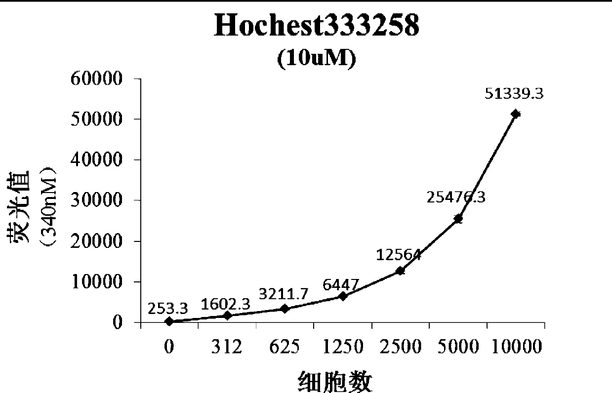
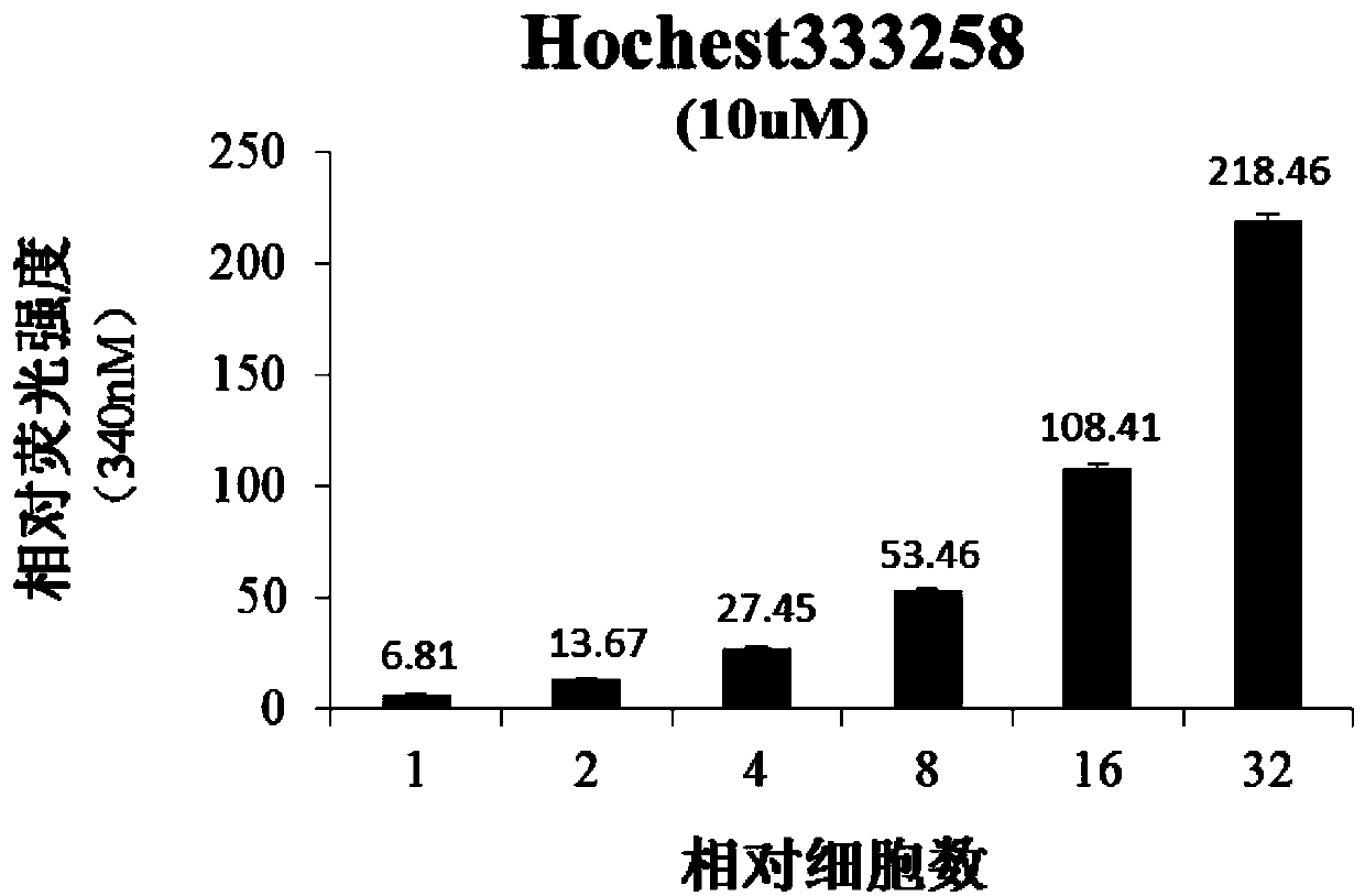
[0036] 2) subculture human non-small cell lung cancer A549 cells into 96-well cell culture plates, count the number of cells and then dilute them to 10000, 5000, 2500, 1250, 625, 312 per well, respectively set up four duplicate wells, Then routinely culture in DMEM medium containing 10% calf serum (humidity 95%, temperature 37° C., 5% CO 2 ) for 24 hours. Then Hochest333258 was added to the cell culture medium to a final concentration of 10 μM. After mixing, place at room temperature for 10 minutes, then discard the cell culture medium, wash off the residual medium with PBS buffer, and then add 100 μL / well of PBS buffer. The wells without cell culture but added with 100 μl / well of PBS buffer were used as blank control, and the fluorescence value was detected at a wavelength of 340 nm using a fluorescent microplate reader.

[0037] Then two independent experiments were carri...

Embodiment 2

[0042] Other cases of tumor cell proliferation detection:

[0043] 1) Subculture human breast cancer MCF-7 cells, human cervical cancer Hela cells, and mouse melanoma B16-F1 cells to 96-well cell culture plates respectively, and after counting the number of cells, doubling according to the method described in Example 1 Dilution, cell culture.

[0044] 2) The cell nuclei were stained with the same concentration of Hochest333258 by the method described in Example 1, and the fluorescence intensity and relative fluorescence intensity of the cells were analyzed.

[0045] 3) The fluorescence value and relative fluorescence intensity of MCF-7, Hela and B16-F1 cells are as follows Figure 4 and 5 shown, where Figure 5 In each group of histograms, the left one is MCF-7 cells, the middle one is Hela cells, and the right one is B16-F1 cells. As the cell number increases, the fluorescence of MCF-7, Hela and B16-F1 cells The values ​​and relative fluorescence intensities also increase...


PUM


Abstract

The invention discloses a tumor cell proliferation detection method based on Hochest333258 and application. The method comprises the following steps: subculturing tumor cells to a cell culture plate,counting cells, then carrying out doubling dilution, so that a gradient series with different cell numbers is obtained, carrying out the conventional culture in a culture medium, then adding liquid containing Hochest333258, uniformly mixing, then standing for a period of time, abandoning a cell culture medium, washing off residual culture medium with PBS buffer liquid, then adding the PBS buffer liquid, finally detecting a fluorescent value, and judging cell proliferation level or survival rate by virtue of fluorescence signal intensity. The method disclosed by the invention is simple and is high in repeatability and accuracy; and the proliferation level of the tumor cells can be effectively detected.

Description

technical field [0001] The invention relates to the field of biomedicine, in particular to a method for detecting cell proliferation based on Hochest333258-labeled tumor cells and its application. Background technique [0002] The level of tumor proliferation is closely related to the degree of malignancy of tumor cells, and it is an important detection index for anti-tumor research and treatment. In the screening and identification of anti-tumor drugs and the research of tumor therapeutic targets, the detection of tumor cell proliferation level is a basic research method. At present, the commonly used method is: use the tumor cell line cultured in vitro as a model, and use MTT method, CCK-8 method, cell counting method or crystal violet staining method to detect after a period of intervention with drug treatment, gene regulation and other conditions, The number of viable tumor cells. Then, the survival rate or inhibition rate of the tumor cells is calculated by comparing ...

Claims


Application Information

Patent Timeline
Patent Type & Authority Applications(China)
IPC IPC(8): G01N21/64G01N33/574
CPCG01N21/6428G01N33/57484G01N2800/7023
Inventor 曹春雨王艳林杨建林王发玲
Owner CHINA THREE GORGES UNIV
Why Eureka
Try Eureka