Cardiac ultrasound imaging method

An ultrasonic imaging method and ultrasonic image technology, applied in the directions of ultrasonic/sonic/infrasonic image/data processing, ultrasonic/sonic/infrasonic diagnosis, ultrasonic/sonic/infrasonic Permian technology, etc., which can solve inaccurate volume calculations, etc. problem, to achieve high operating efficiency, enhance the edge area, and improve the effect of segmentation

Active Publication Date: 2018-05-11
CHISON MEDICAL TECH CO LTD
View PDF6 Cites 14 Cited by
  • Summary
  • Abstract
  • Description
  • Claims
  • Application Information

AI Technical Summary

Problems solved by technology

[0004] The purpose of the present invention is to overcome the deficiencies in the prior art and provide a cardiac ultrasound imaging method, which solves the problem of automatically segmenting the contour of the left ventricle during the heart measurement process and prevents manual designation of the left ventricle segmentation point during the measurement process. Circumstances that may result in inaccurate volume calculations

Method used


Image

  • Cardiac ultrasound imaging method
  • Cardiac ultrasound imaging method
  • Cardiac ultrasound imaging method

Examples


Embodiment Construction

[0039] The present invention will be further described below in conjunction with drawings and embodiments.

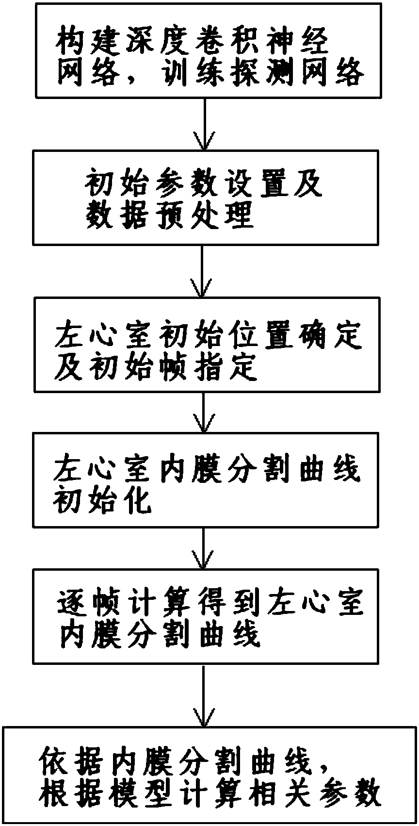
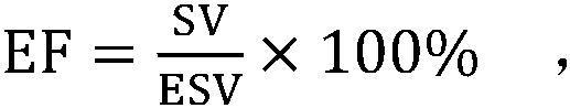
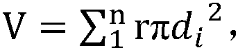
[0040] Such as figure 1 As shown, the present invention discloses a cardiac ultrasound imaging method, comprising the following steps: 1) constructing a deep convolutional neural network and training a detection network; 2) initial parameter setting and data preprocessing; 3) determination of the initial position of the left ventricle and initial Frame designation; 4) initialization of the left ventricular endocardial segmentation curve; 5) calculation of the left ventricular endocardial segmentation curve frame by frame; 6) calculation of left ventricular volume, ejection fraction and other related parameters based on the intima segmentation curve and the model.

[0041] In order to solve the problem of automatically segmenting the contour of the left ventricle during the heart measurement process and prevent the inaccurate volume calculation that may be caused by manu...


PUM


Abstract

The invention discloses a cardiac ultrasound imaging method, and aims at solving the problem of automatically segmenting of a left ventricle contour during a cardiac measurement process, and preventing the situation of inaccurate volume calculation which might be caused by manually specifying left ventricular segmentation points in the measurement process. Compared with a traditional left ventricular segmentation algorithm, a large number of images of the position of a left ventricle manually marked by a doctor are used to train a deep convolutional neural network, the position of the left ventricle can be accurately identified by the acquired detection network, and a level set algorithm can be performed within a detection position area to obtain a segmentation curve of a left ventricularendocardium; compared with a traditional algorithm, acquired results are more robust, the operation efficiency is higher. According to the cardiac ultrasound imaging method, a manual enhancement manner is adopted to enhance edge areas to improve a segmentation effect aiming at the situation of poor image quality of ultrasound mapping.

Description

technical field [0001] The invention relates to the field of ultrasonic image processing, in particular to a cardiac ultrasonic imaging method. Background technique [0002] Two-dimensional real-time echocardiography is a non-invasive diagnostic and monitoring technique widely used in clinical diagnosis and treatment of cardiovascular diseases. Although accurate delineation of the intimal boundary of the left ventricle during diastole and systole can provide important cardiac function parameters for clinical practice, However, due to the quality of ultrasound images, it has always been very difficult to delineate ultrasound boundaries fully or even semi-automatically. [0003] At present, there are some left ventricle segmentation algorithms that use the level set method to obtain the segmentation curve of the left ventricle endocardium. The initial level set generated by the algorithm has low accuracy. Contents of the invention [0004] The purpose of the present invent...

Claims


Application Information

Patent Timeline
Patent Type & Authority Applications(China)
IPC IPC(8): A61B8/08A61B8/00
CPCA61B8/0883A61B8/5207A61B8/5223
Inventor 甘从贵赵明昌陆坚
Owner CHISON MEDICAL TECH CO LTD
Why Eureka
Try Eureka