Transperineal needle guidance

A technology of guiding holes and guiding components, applied in the directions of catheters, trocars, parts of surgical instruments, etc., can solve problems such as image movement

Active Publication Date: 2018-06-08
ZMK MEDICAL TECH INC
View PDF17 Cites 6 Cited by
  • Summary
  • Abstract
  • Description
  • Claims
  • Application Information

AI Technical Summary

Problems solved by technology

However, where it is desired to obtain multiple 2D images for 3-D image generation, manual manipulation of the device can cause significant motion between images

Method used


Image

  • Transperineal needle guidance
  • Transperineal needle guidance
  • Transperineal needle guidance

Examples


Embodiment Construction

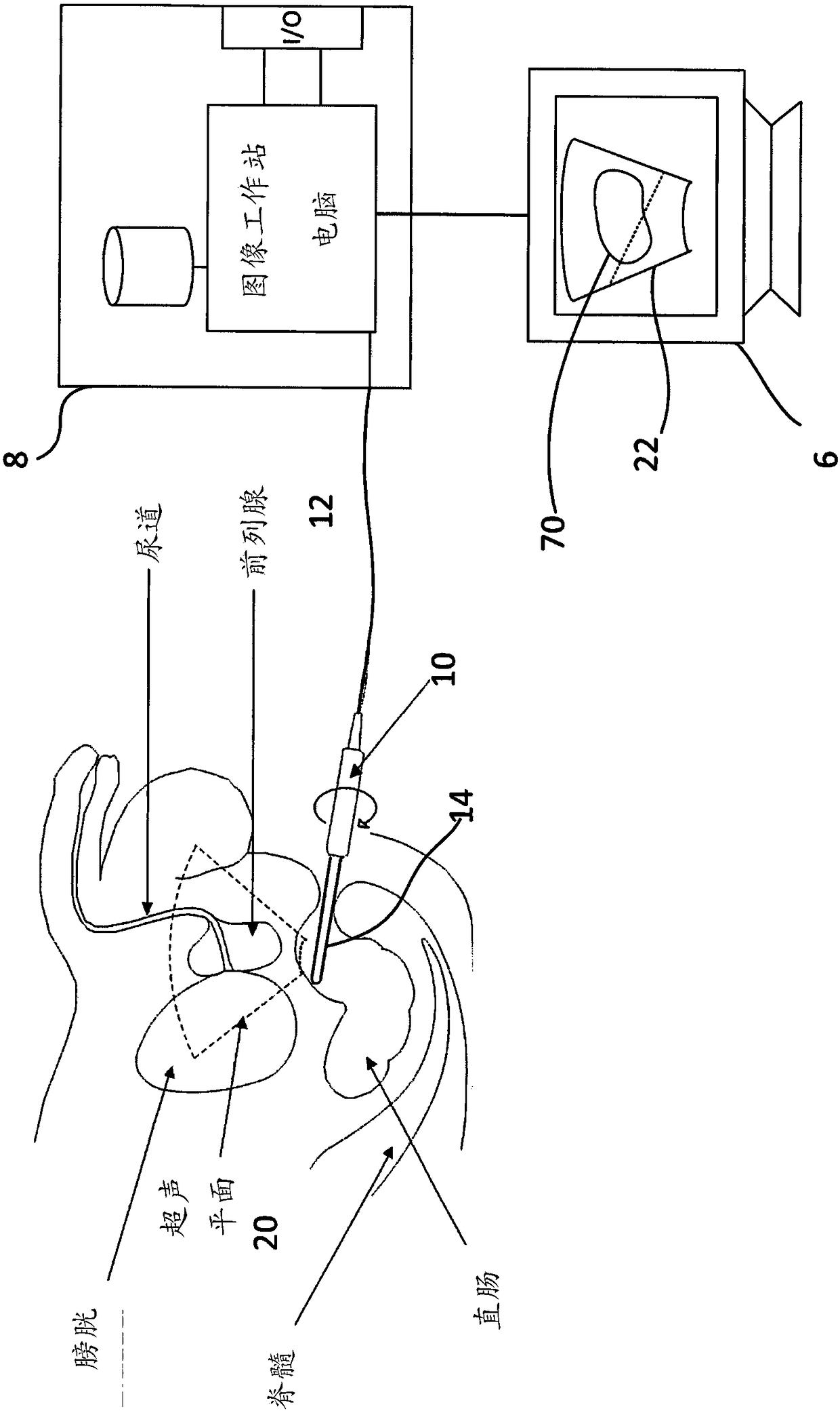
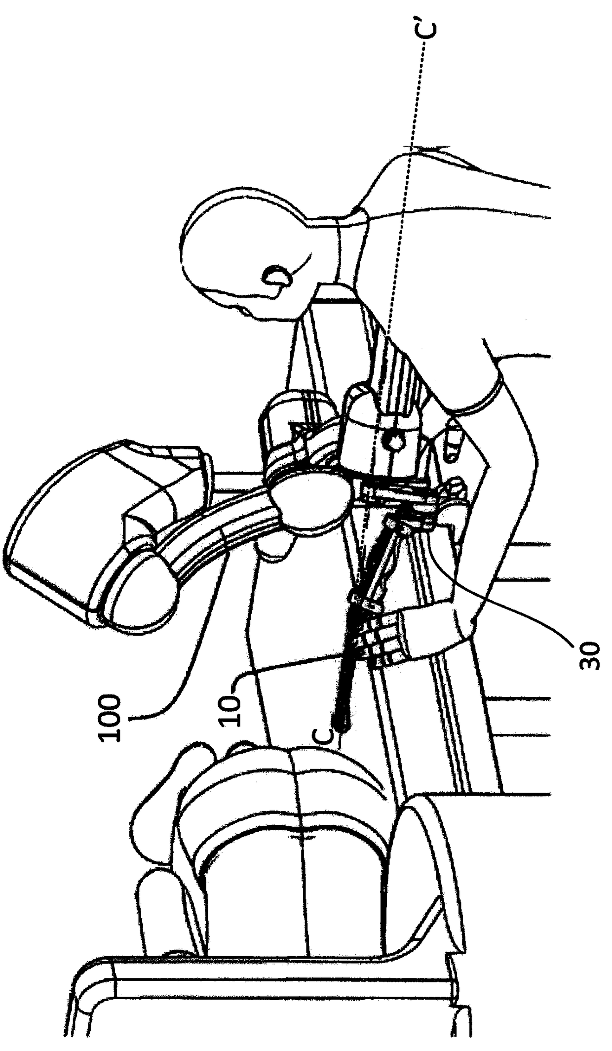
[0025] Reference is now made to the accompanying drawings, which help illustrate various features of the invention. While the invention is first described with respect to transrectal ultrasound imaging, biopsy and therapy for prostate imaging, it should be understood that some aspects of the invention may be applicable to other medical imaging applications. In this regard, the following description is for purposes of illustration and description.

[0026] The disclosed systems and methods facilitate obtaining medical images and / or performing medical procedures. One embodiment provides a combined medical imaging device holder (eg, probe holder) and guide assembly. The guide assembly holds the supported placement element (e.g., needle, trocar, treatment device, etc.) in the image area / plane of the imaging device (e.g., the two-dimensional image plane of the ultrasound probe) held by the probe holder, the The placement element is adapted for insertion into patient tissue. The ...


PUM


Abstract

A combined imaging probe holder and interventional needle guide assembly is provided. The imaging probe holder supports an imaging device (e.g., ultrasound probe) in a known relationship with a biopsy / treatment device holder such that a delivery element or interventional needle is constrained within an imaging field of the imaging device. Constraining the biopsy / treatment device within the imagingfield allows for real-time monitoring of the biopsy / treatment device during insertion of the interventional needle into patient tissue. In addition, the biopsy / treatment device holder is angularly positionable relative to the image field to allow advancement of the delivery element to any desired location within the imaging field.

Description

[0001] cross reference [0002] This application claims the benefit of the filing date of U.S. Provisional Application No. 62 / 189,352, which was filed July 7, 2015, the entire contents of which are hereby incorporated by reference in their entirety. technical field [0003] The present invention relates to systems, methods and devices (utilities) allowing tracking and guidance of interventional needles or needle applicators (biopsy needles, treatment devices) through the rectum or perineum of a patient. More specifically, the present invention relates to a utility that allows guiding and holding a medical imaging instrument and a needle guiding device in a fixed positional relationship to each other for guiding any interventional needle passing through a patient's rectum or perineum in real time to a desired target location . Background technique [0004] Physicians and other medical professionals often use medical imaging equipment to perform non-invasive examinations. Th...

Claims


Application Information

Patent Timeline
Patent Type & Authority Applications(China)
IPC IPC(8): A61B8/08A61B8/12A61B10/02
CPCA61B8/0841A61B8/12A61B8/4209A61B8/4218A61B8/4263A61B8/4455A61B8/463A61B8/466A61B8/483A61B8/5246A61B17/3403A61B18/1477A61B2017/00274A61B2017/3405A61B2017/3411A61B2017/3413A61B2018/00547A61B2018/0293A61N5/1027A61N5/1049A61N2005/1012A61N2005/1058A61B90/11A61B2034/2059A61B2090/365A61B2090/3782A61B2090/3784
Inventor 许光耀R.文卡塔拉曼S.萨卡尔M.艾哈迈迪
Owner ZMK MEDICAL TECH INC
Why Eureka
Try Eureka