Image guidance system

a technology of image guidance and image, applied in the field of medical image guided interventions, can solve the problems of increasing the time in the operation room, increasing the cost, and limited availability of clean operating room, so as to improve the clinical workflow and facilitate the efficient use of biopsy sample analysis

Active Publication Date: 2018-06-21
KONINK PHILIPSN V
View PDF3 Cites 5 Cited by
  • Summary
  • Abstract
  • Description
  • Claims
  • Application Information

AI Technical Summary

Benefits of technology

[0019]The treatment plan could also be created during the combined biopsy and treatment procedure. Hereby, optionally also the biopsy result could be used for the creation of the treatment plan. Also before a biopsy result is retrieved multiple treatment plans could be created, which are dependent on the biopsy outcome. In this way after the biopsy result is retrieved, the user can select the treatment plan that best matches the biopsy result. In this way, waiting time for biopsy sample analysis can be efficiently used. In the above described cases the ultrasound image could be used as planning image. The image guidance means could comprise delineation means, such that a user could delineate regions of interest, like the target and one ...

Problems solved by technology

The availability of such clean operating rooms is limited.
Executing this patient...

Method used


Image

  • Image guidance system
  • Image guidance system
  • Image guidance system

Examples


Embodiment Construction

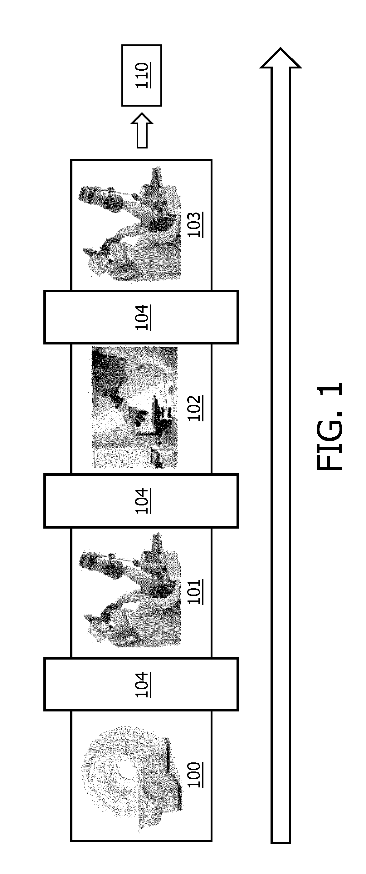
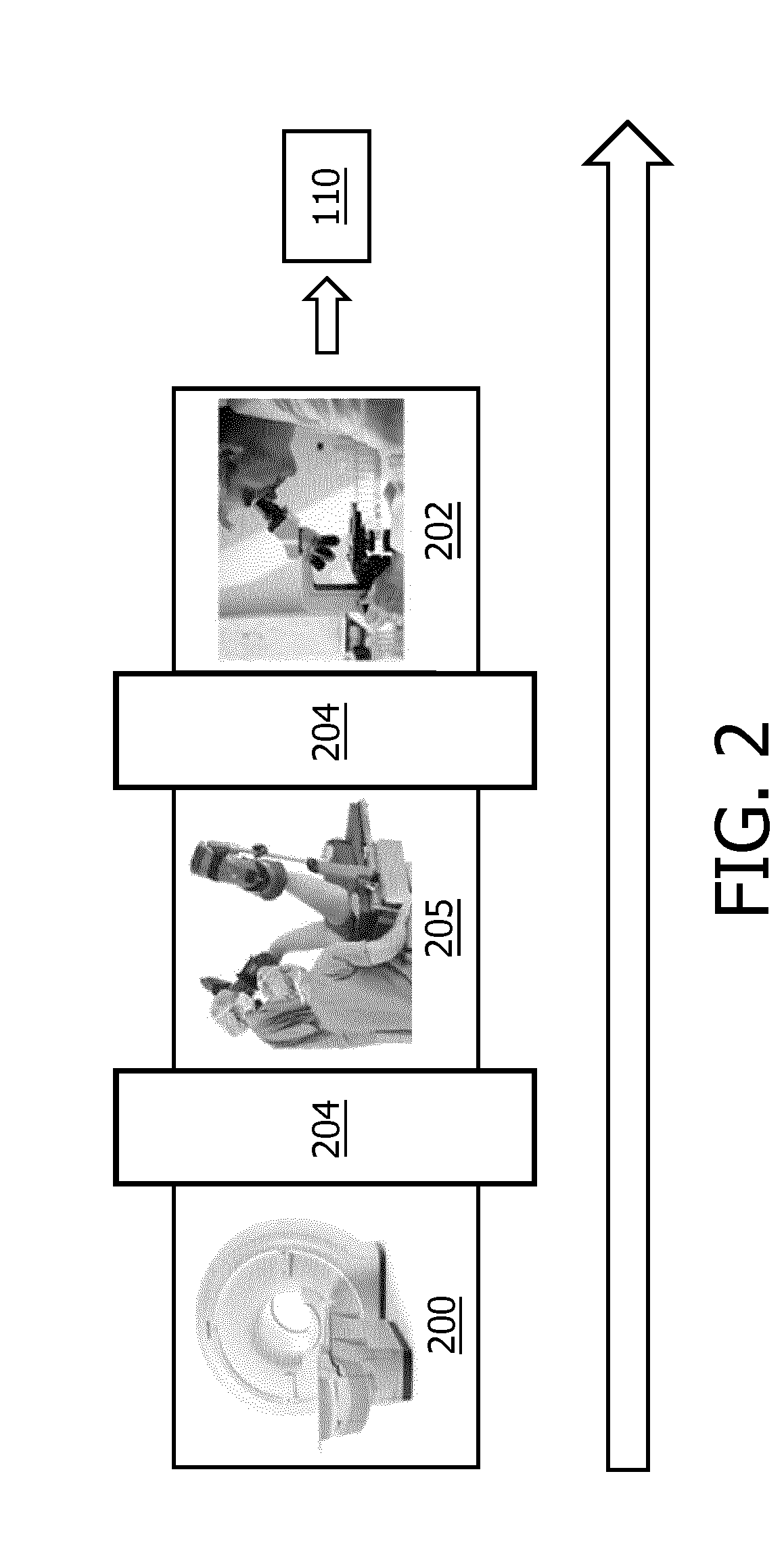
[0027]FIG. 1 shows an illustration of a clinical workflow which is known in the art. First a medical image is acquired 100 from the region of interest in the subject of interest. In this case the region of interest is a prostate. The acquired medical image is an MRI image. A physician determines regions (predetermined end locations) that are suspicious for comprising tumor tissue based on the MRI image. This will usually result in a delay of several days 104. After the delay 104 the patient has to come back to the hospital for the biopsy procedure 101. This biopsy could be performed under ultrasound guidance. The MRI image will be registered to the ultrasound image, such that that the predetermined end locations can be translated to a coordinate system of the ultrasound imaging system. A biopsy will be taken from these predetermined end locations. Then the patient will be send back home. The biopsy samples will be send to a pathology department, which will cause a further delay 104 ...


PUM


Abstract

It is an object of the invention to provide for a system that can be used to improve a cancer related clinical workflow compared to the current cancer related clinical workflow. This object is achieved by an image guided system configured for supporting a combined biopsy and treatment procedure using image guidance to bring a needle from a defined location to a predetermined end location in a subject of interest. The image guidance system comprises a medical imaging system (580) configured for acquisition of medical images of a region of interest (403) in the subject of interest, wherein the medical images are used to determine a current position of the predetermined end location (404). The image guidance system further comprises needle guidance system (402, 403) configured for defining a needle position in space and thereby defining the defined location (405, 406) and configured for guiding a biopsy and treatment needle into the region of interest. The image guidance system also comprises a user interface system (530) configured for supporting both the biopsy and treatment procedure, such that a patient setup needs to be executed only once. The user interface system (530) comprises a calibration unit (540) for aligning the needle guidance system (402, 403) with the medical images.

Description

FIELD OF THE INVENTION[0001]The invention relates to a system in the field of medical image guided interventions.BACKGROUND OF THE INVENTION[0002]Amongst others, diagnosis of cancer is performed based on tissue samples obtained in a biopsy procedure. These biopsy procedures are often performed systematically, taking a number of biopsy samples from a standardized set of locations inside the prostate. With the availability of fusion-guided biopsy systems, this is now shifting to a more targeted approach in which a suspicious lesion as shown on magnetic resonance imaging (MRI) is being sampled. If a patient has a biopsy proven tumor, this patient is often treated, e.g. by means of radiotherapy.[0003]At present, altogether, a complete clinical flow from MRI, via fusion guided biopsy and pathology to treatment, typically takes 2-4 weeks in most hospitals. During this workflow first an MRI image may be acquired on which suspicious tissue may be identified. The MRI image could be used duri...

Claims


Application Information

Patent Timeline
IPC IPC(8): A61B10/02A61B17/34
CPCA61B10/0241A61B17/3403A61B2017/3413A61B2017/3411A61B34/10A61B34/20A61B90/11A61B2034/107A61B2034/2051A61B2090/364A61B2090/3782
Inventor HAUTVAST, GUILLAUME LEOPOLD THEODORUS FREDERIKKUSTRA, JACEK LUKASZ
Owner KONINK PHILIPSN V
Why Eureka
Try Eureka