Visualization of three-dimensional image data on a two-dimensional image

a two-dimensional image and image data technology, applied in the field of medical imaging, can solve the problems of inability to plan, perform, analyze the effectiveness of the procedure with sufficient simplicity, accuracy, and difficulty in reaching the target point from the chosen insertion point using the device, and achieve the effect of easy visualization

Active Publication Date: 2020-04-23
CANON USA
View PDF0 Cites 5 Cited by
  • Summary
  • Abstract
  • Description
  • Claims
  • Application Information

AI Technical Summary

Benefits of technology

[0010]Accordingly, it is a broad object of the present disclosure to provide devices, systems, and methods to allow a clinician to easily visualize 3D images on a 2D display to provide planning, performance, and evaluation techniques.
[0015]In accordance with at least another aspect of the present disclosure, the planning, performance, and evaluation technique(s) systems and methods discussed herein may be employed to reduce the cost of at least one of manufacture and maintenance of planning, performance, and evaluation devices, systems and storage mediums by reducing or minimizing a number of components therein to cut down cost.

Problems solved by technology

Thus, when viewing a two-dimensional image on the screen, it can be difficult to visualize and understand the relationship between the target lesion, the skin entry point, and any critical structures or other features that could affect the intervention.
Thus, in conventional systems, there is limited ability to plan, perform, and analyze the effectiveness of the procedure with sufficient simplicity, accuracy, no provision for simulating the device placement in the imaging software during planning for evaluating target reachability.
Thus, before the needle guidance device is placed on the patient, imaged and registered to the image space, it is difficult to be certain that the target point is reachable from the chosen insertion point using the device.
While U.S. Pat. No. 9,710,146; U.S. Pat. Pub. 2011 / 0007071 and others have provided systems and methods for image display control that supports a user in selecting an image of interested from among a three-dimensional data set, it does not allow for a simple and intuitive way of planning and carrying out minimally invasive procedures for needle guidance.

Method used


Image

  • Visualization of three-dimensional image data on a two-dimensional image
  • Visualization of three-dimensional image data on a two-dimensional image
  • Visualization of three-dimensional image data on a two-dimensional image

Examples


Embodiment Construction

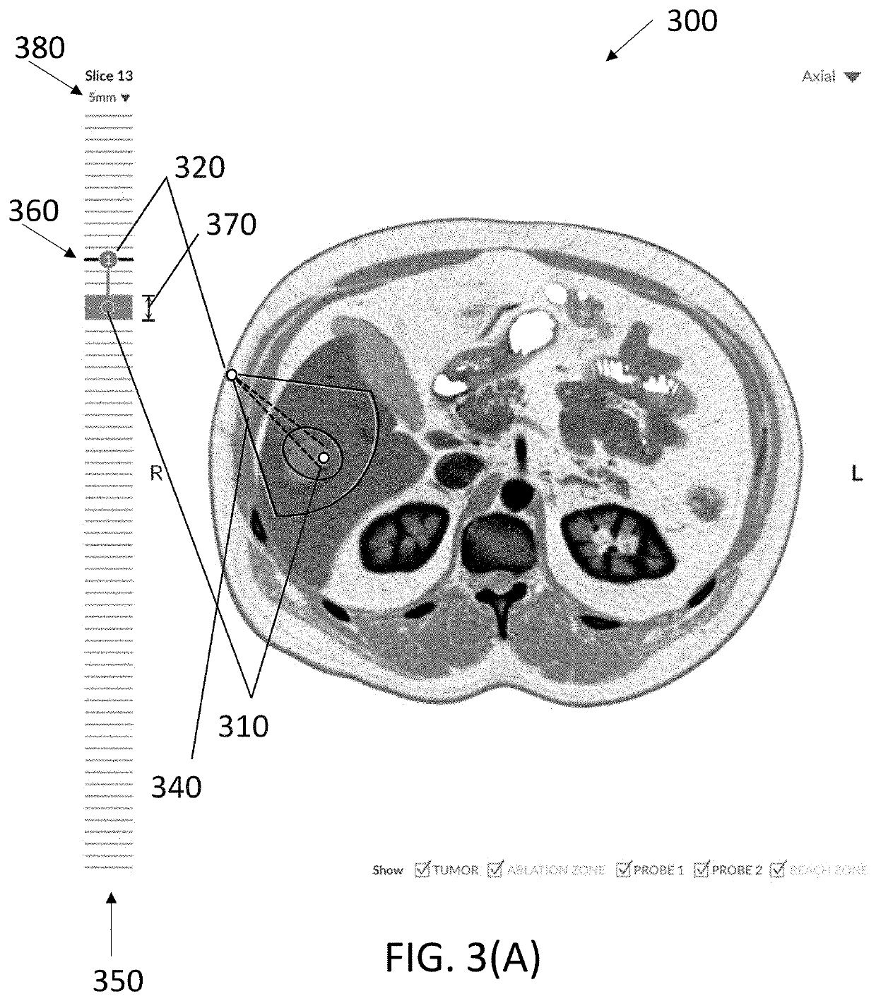
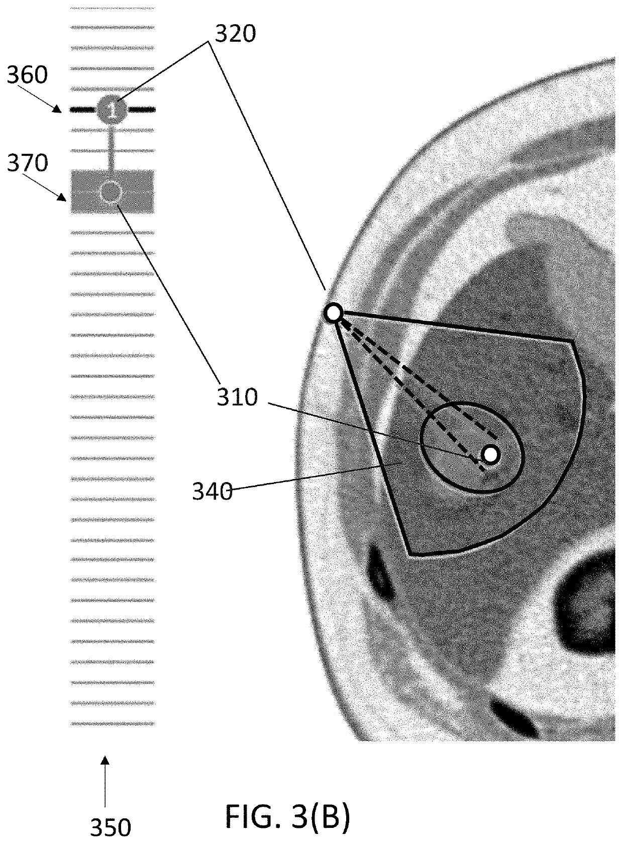
[0026]Exemplary embodiments are described below with reference to the drawings. The present invention provides for improved visualization of image date where the image date is two-dimensional slices of three-dimensional (3D) image set. While many clinicians are comfortable paging through the various slices to obtain an understanding of the region of interest and surrounding tissue, as the planning and / or procedures become more complicated, the ability to visualize information from the 3D image set when viewing a two-dimensional (2D) image become important.

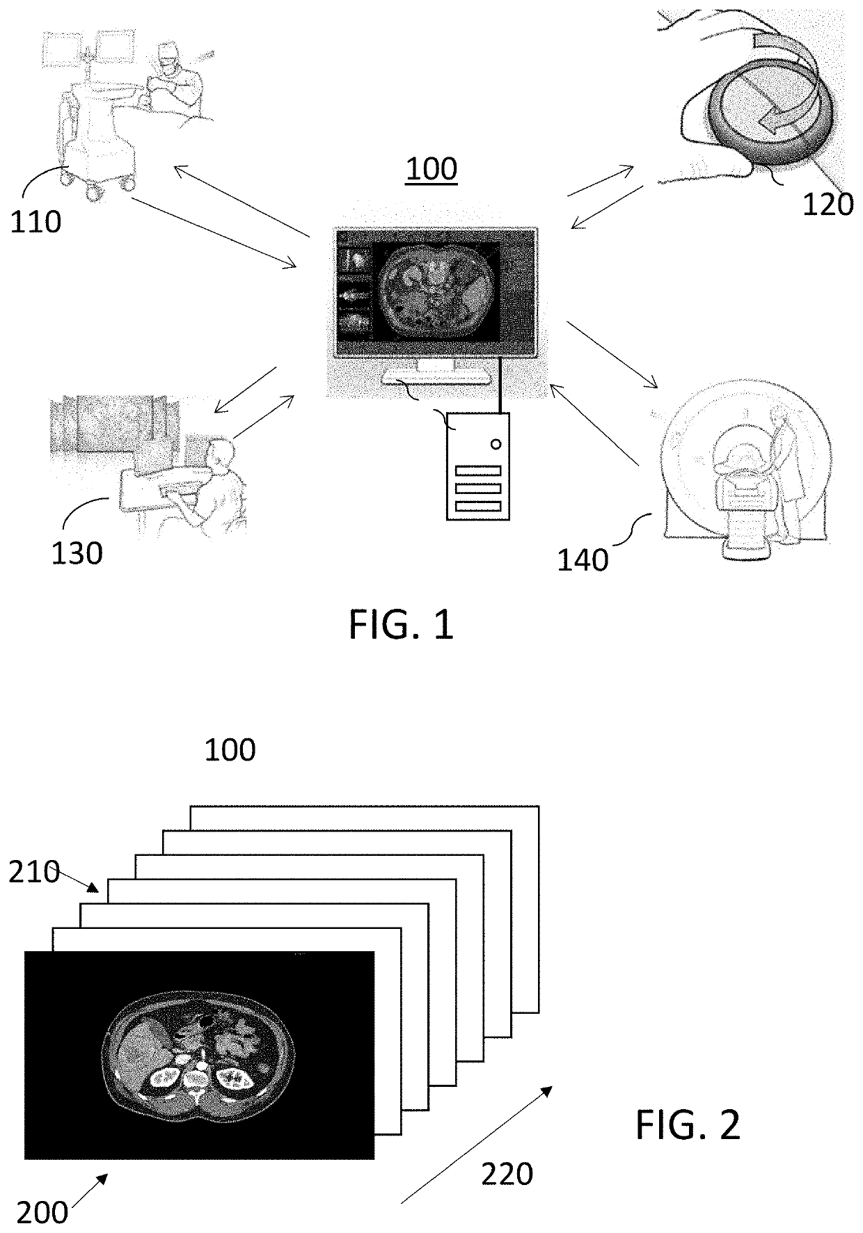
[0027]For example, in some embodiments, a clinician will plan, perform, and / or evaluate performance. FIG. 1 illustrates a system with displayed image data 100, a model system and cart 110, a device model 120, and registered device model 130 that can be overlaid on the displayed image data 100. In some embodiments, the planning image data 100 is a three-dimensional image set obtained from an imaging system (CT, MRI, etc.) 140. In so...


PUM


Abstract

The present disclosure relates generally to medical imaging and, more particularly to systems, methods, and devices for planning and carrying out minimally invasive procedures using external devices for needle guidance and the display and manipulation of the image set when planning and performing the procedure.

Description

CROSS-REFERENCE TO RELATED APPLICATION(S)[0001]This application relates, and claims priority, to U.S. Patent Application Ser. No. 62 / 748,168, filed Oct. 19, 2018, the entire disclosure of which is incorporated by reference herein in its entirety.FIELD OF THE INVENTION[0002]The present disclosure relates generally to medical imaging and, more particularly to systems, methods, and devices for planning and carrying out minimally invasive procedures using external devices for needle guidance.BACKGROUND OF THE INVENTION[0003]Minimally invasive medical procedures are becoming increasingly popular in the medical community due to shortened hospital stays and improved quality of life for the patient. For example, in the field of interventional oncology, percutaneous ablations are often preferred over surgical resection due to the minimally invasive nature of the procedure and thus shortened patient recovery period.[0004]Medical images assist physicians in planning, performing, and post-opera...

Claims


Application Information

Patent Timeline
Patent Type & Authority Applications(United States)
IPC IPC(8): A61B34/10G06T7/00A61B34/00A61B34/20
CPCA61B34/25G06T2207/10081A61B2034/2065G06T7/0014A61B34/10A61B34/20A61B2034/107G06T2207/10132G06T2207/10088A61B2034/104A61B2034/2051A61B2034/102A61B2034/108A61B2090/363A61B2017/00973A61B17/3403A61B2034/2059G06T19/003G06F3/04845
Inventor NAKAMURA, HITOSHIDANIELS, BARRETBONILLAS VACA, ANTONIOTHURROTT, CHRISTOPHER WAYNERUDOLPH, MORGAN CARLSON
Owner CANON USA
Why Eureka
Try Eureka