Colposcopy image-based cervical cancer detection method, device and equipment and medium

A detection method and cervical cancer technology, applied in the field of medical image processing, can solve the problems of missing information of small objects and poor effect, and achieve the effect of reducing constraints and promoting cervical cancer and precancerous lesions

Inactive Publication Date: 2018-09-07
姚书忠 +3
View PDF5 Cites 29 Cited by
  • Summary
  • Abstract
  • Description
  • Claims
  • Application Information

AI Technical Summary

Problems solved by technology

However, the effect on segmenting small objects is poor. As the network level deepens, although a deeper network can better fit the sample distribution, the lack of information on small objects is more serious.

Method used


Image

  • Colposcopy image-based cervical cancer detection method, device and equipment and medium
  • Colposcopy image-based cervical cancer detection method, device and equipment and medium
  • Colposcopy image-based cervical cancer detection method, device and equipment and medium

Examples


no. 2 example

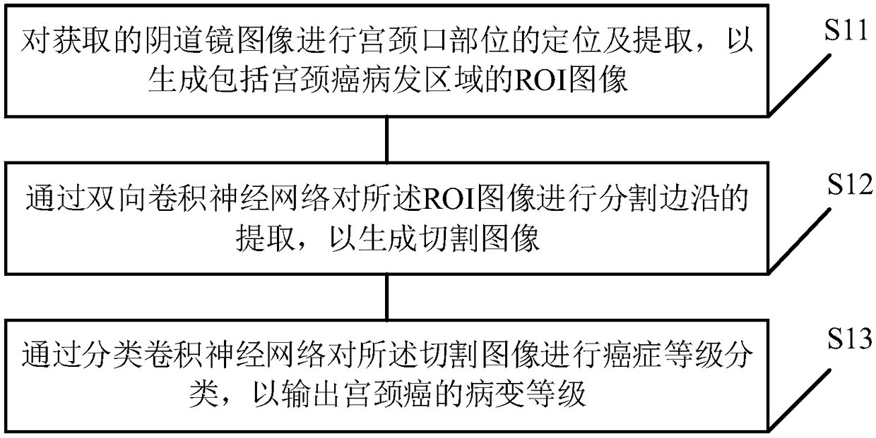
[0068] see Figure 8 , On the basis of the first embodiment of the present invention, the training steps of the cervical cancer detection model based on the bidirectional convolutional neural network include:

[0069] S21. Perform preprocessing on the acquired colposcope sample image to generate a first sample.

[0070] In the embodiment of the present invention, specifically, in order to ensure the reliability of the image quality of the colposcope sample, data enhancement is performed on the original colposcope sample image under the premise of ensuring that the contrast of the colposcope sample image is not changed. The enhancement method is mainly as follows: Translate, flip, add noise, etc. Flip is to rotate the image in 3 directions and the mirror image of the original image; the added noise is common Gaussian noise to form the first sample after preprocessing.

[0071] S22. Perform sample expansion on the first sample according to the adversarial generative network to...


PUM


Abstract

The invention discloses a colposcopy image-based cervical cancer detection method, device and terminal equipment and a computer readable storage medium. The method comprises the following steps of: ina bidirectional convolutional neural network-based cervical cancer detection model, positioning and extracting a cervical opening position of an obtained colposcopy image so as to generate an ROI image comprising a cervical cancer disease area; extracting a cutting edge of the ROI image through a bidirectional convolutional neural network so as to generate a cut image; and carrying out cancer grade classification on the cut image through a classification convolutional neural network so as to output a cervical cancer lesion grade and then rapidly and correctly obtain a cervical cancer detection result. According to the method, doctors that are lack of experiences can be helped to rapidly judge diseased regions, discover atypical diseased regions and judge the disease degrees and sampling regions, so that help and auxo-action are provided for discovering cervical cancer and precancerous lesions.

Description

technical field [0001] The present invention relates to the field of medical image processing, in particular to a colposcope image-based cervical cancer detection method, device, terminal equipment and computer-readable storage medium. Background technique [0002] Image segmentation algorithm already exists in the field of traditional digital image processing, mainly using the threshold segmentation method and its various variants. On the basis of the image segmentation algorithm, the image detection algorithm is extended, but they have different levels of image processing. The detection is mainly aimed at the image area level, while the segmentation needs to process each pixel, and the image processing is more delicate. In the field of deep learning, there are more and more segmentation algorithms based on convolutional networks, and the most widely used is the fully convolutional network (FCN). [0003] Existing cervical cancer pre-cancer screening is based on the "three...

Claims


Application Information

Patent Timeline
Patent Type & Authority Applications(China)
IPC IPC(8): G06T7/00G06T7/12
CPCG06T7/0012G06T2207/20081G06T2207/20084G06T2207/30096G06T7/12
Inventor 姚书忠刘文彬崔淑芬张丽贞
Owner 姚书忠
Why Eureka
Try Eureka