Segmentation method for esophagus cancer in chest CT images

A CT image, esophageal cancer technology, applied in the field of medical image processing, can solve the problems of low contrast, small occupation ratio, difficult to obtain esophageal cancer area, etc., and achieve the effect of fast segmentation, small model size and high accuracy

Inactive Publication Date: 2018-09-28
GUILIN UNIV OF ELECTRONIC TECH +1
View PDF2 Cites 19 Cited by
  • Summary
  • Abstract
  • Description
  • Claims
  • Application Information

AI Technical Summary

Problems solved by technology

Since the esophagus only accounts for a small part of the body on CT slices and is closely related to other related organs, the proportion of the area occupied by other tissues and organs is small, and the contrast with o

Method used


Image

  • Segmentation method for esophagus cancer in chest CT images
  • Segmentation method for esophagus cancer in chest CT images

Examples


Embodiment

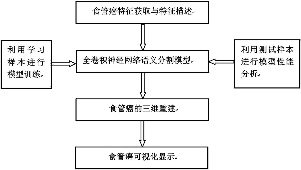
[0020] Such as figure 1 As shown, a method for segmenting esophageal cancer in a chest CT image specifically includes the following steps:

[0021] 1) Select multiple groups of CT images containing esophageal cancer, and use the CT images containing esophageal cancer as training samples.

[0022] 2) Preprocess the CT images selected in step 1), obtain the features of esophageal cancer, and perform feature description; specifically, from the DICOM images of chest CT of each layer, convert them according to the window width and window level A bitmap is formed, and the CT image is cropped, and it is cropped into an image of 80×80 pixels, and the esophagus is included in the cropped image, and the cropped image is used as the feature input of the full convolutional neural network.

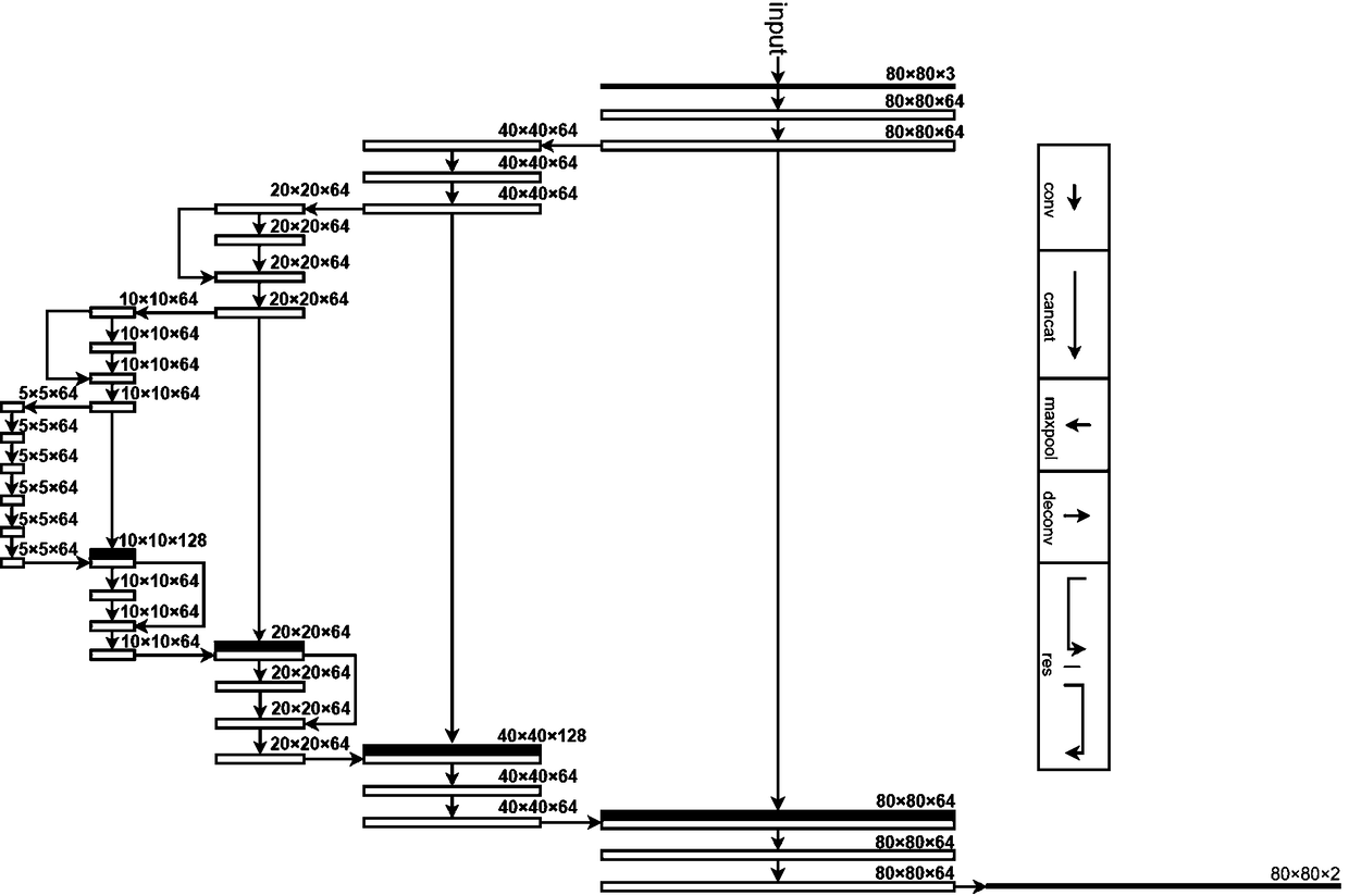
[0023] 3) Establish a semantic segmentation model of esophageal cancer based on the full convolutional neural network, and use the features of esophageal cancer described in step 2) as the feature inp...


PUM


Abstract

The invention discloses a segmentation method for esophagus cancer in chest CT images. According to the method, first, multiple groups of CT images containing the esophagus cancer are selected, and the CT images containing the esophagus cancer are used as training samples; the selected CT images are preprocessed, esophagus cancer features are acquired, and the images obtained after feature description is performed are used as training data; an esophagus cancer semantic segmentation model based on a full-convolution neural network is established, described esophagus cancer features are used asfeature input of the full-convolution neural network to serve as learning samples for training, and an esophagus cancer segmentation network model is obtained; three-dimensional reconstruction is performed on the esophagus cancer, wherein three-dimensional reconstruction and analysis are performed on an esophagus cancer segmentation result obtained through the obtained esophagus cancer segmentation network model, and esophagus cancer image omics parameters under a three-dimensional space are obtained; and the obtained esophagus cancer image omics parameters are visually displayed. The method is small in model scale, high in speed and high in accuracy.

Description

technical field [0001] The invention relates to the technical field of medical image processing, in particular to a method for segmenting esophageal cancer in chest CT images. Background technique [0002] With the continuous development of computer technology, medical imaging is also developing rapidly, so that more and more medical data can be processed and judged by computer, which improves the probability of successful disease diagnosis and treatment. In the diagnosis and detection of esophageal cancer, in addition to endoscopy, computerized tomography (CT) is commonly used. CT images can provide continuous slice grayscale images of a certain part of the human body, and can show the interior of the human body in detail. Therefore, the radiomics method based on CT image processing is often used in the diagnosis and prognosis of esophageal cancer. In the computer-aided diagnosis system, accurate segmentation of CT scan images of esophageal cancer is a key technology for s...

Claims


Application Information

Patent Timeline
IPC IPC(8): G06T7/00G06T7/11G06T17/20
CPCG06T7/0012G06T17/20G06T2207/10081G06T2207/20081G06T2207/20084G06T2207/20132G06T2207/30096G06T7/11
Inventor 陈树超陈洪波刘立志黎浩江徐绍凯傅嘉文朱志华
Owner GUILIN UNIV OF ELECTRONIC TECH
Why Eureka
Try Eureka