
Hysterosalpingography tube

A technology for fallopian tubes and uterine cavity, which is applied in the field of hysterosalpingography tube for contrast-enhanced ultrasound and selective salpingography. Regurgitation, high surgical reliability, and improved diagnostic accuracy

Pending Publication Date: 2018-10-09
ZHEJIANG PROVINCIAL PEOPLES HOSPITAL
View PDF8 Cites 6 Cited by
  • Summary
  • Abstract
  • Description
  • Claims
  • Application Information

AI Technical Summary

Problems solved by technology

One type is hard catheter: its tapered head does not enter the uterine cavity, which reduces the discomfort of patients, but in the operation of patients with cervical relaxation, there are cervical insufficiency, contrast fluid reflux, and unsatisfactory images; secondly, its hard rod During vaginal contrast-enhanced ultrasound, the rotation of the ultrasound probe is greatly hindered, which interferes with the detection accuracy and increases the patient's pain. In addition, in order to prevent contrast fluid reflux imagination, it is often necessary for medical staff to hold the catheter to fix and adjust the direction, which increases manpower. Material resources, but also prolong the operation time
The other is a soft tubing. After it is placed into the internal os of the cervix, a balloon is used to seal the internal os of the cervix to prevent reflux of the contrast fluid. In order to fully seal the internal os of the cervix, it is often necessary to inject at least 3ml of gas or liquid, and the balloon is inflated. The posterior volume is large, and its image will hinder the development of uterine cavity lesions; and the inflated balloon will stimulate the cervix, induce vagal reflex, and cause nausea, vomiting and other discomfort during the operation in most patients, which will affect the operation.
[0003] Both of the above two existing contrast tubes have the defect of false positive results due to short-term spasm of the fallopian tube orifice, resulting in subsequent misjudged results and increasing the probability of further interventional treatment for patients

Method used


Image

  • Hysterosalpingography tube
  • Hysterosalpingography tube
  • Hysterosalpingography tube

Examples


Embodiment Construction

[0025] The present invention will be specifically introduced below in conjunction with the accompanying drawings and specific embodiments.

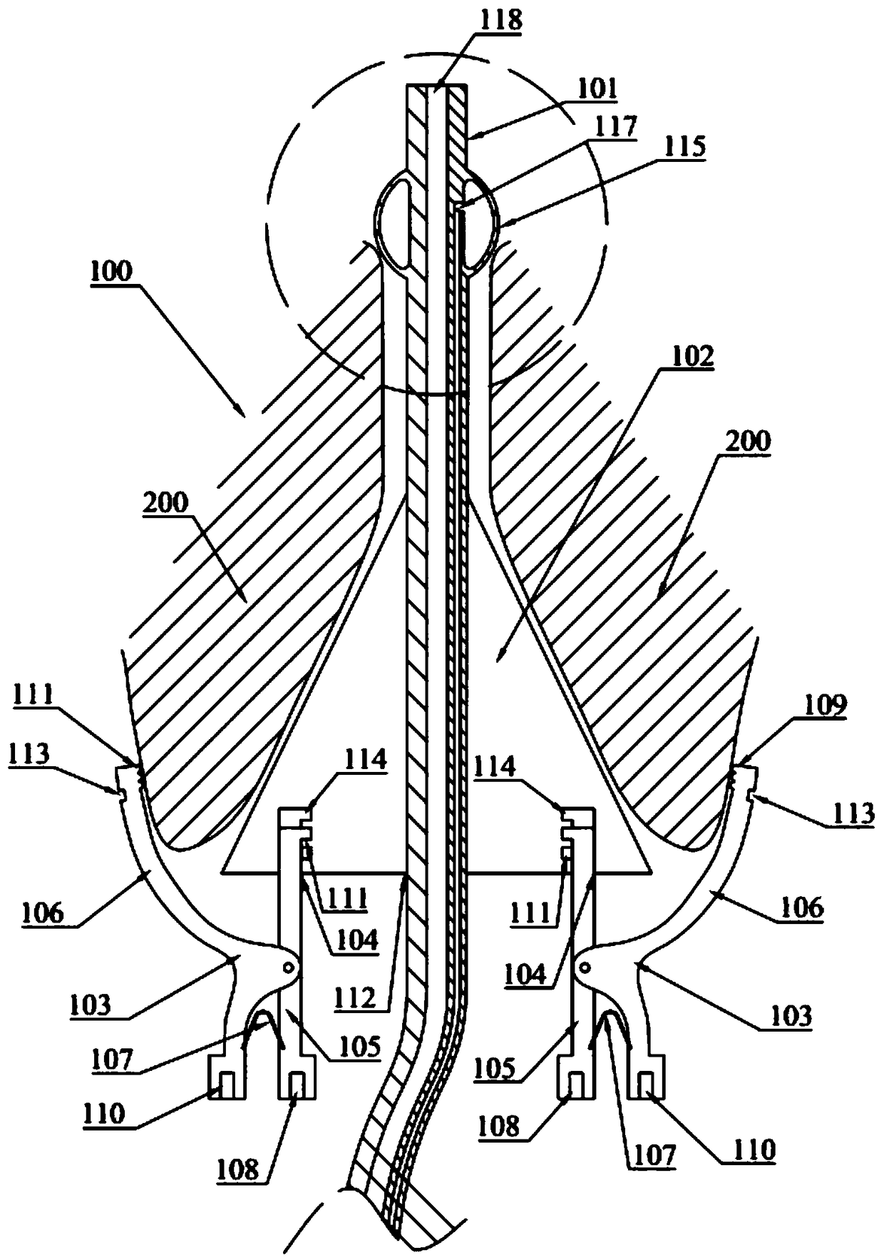
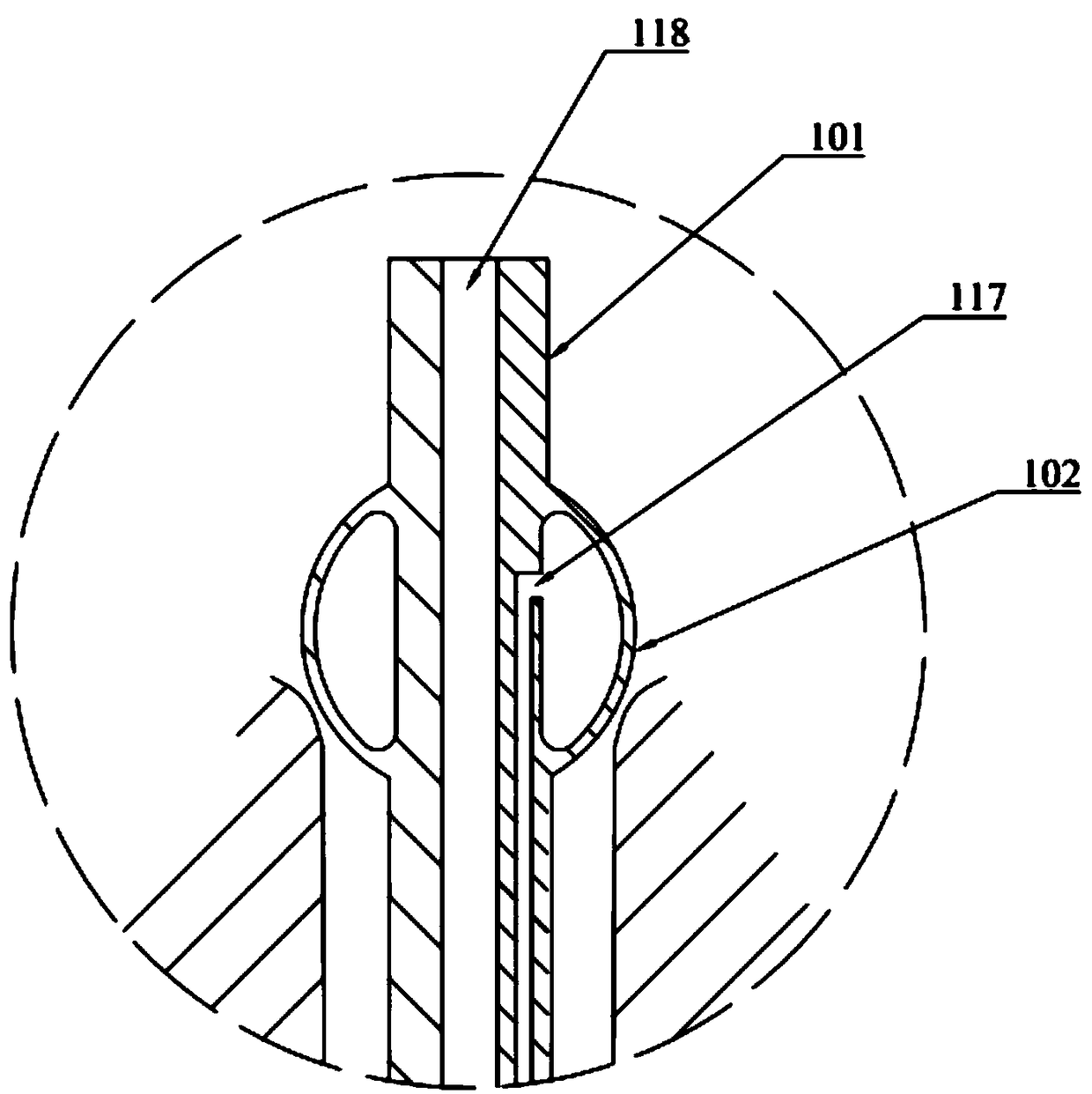
[0026] Such as Figure 1 to Figure 4 As shown, a hysterosalpingography tube 100 includes: a soft tubing 101, a tapered head 102 for closing the external os of the cervix, two clamps 103 for clamping the cervix 200, and a tubal interventional catheter for directional input of contrast fluid into the fallopian tubes 119; one end of the soft tube 101 passes through the conical head 102 and forms a balloon 115 that is inflated or filled with liquid to close the internal os of the cervix; the soft tube 101 is formed with a guide channel 118 for the fallopian tube interventional catheter 119 to penetrate and for Balloon 115 is inflated or liquid-filled filling channel 117; two fixing clips 103 are respectively connected to the two sides of conical head 102; conical head 102 is formed with slot 104; 106 and spring leaf 107; Inner clamp arm 105 ...


PUM


Abstract

The invention discloses a hysterosalpingography tube. The hysterosalpingography tube comprises a soft tube, a conical head for closing the outer opening of the cervix, two fixing clamps for clamping the cervix, and a fallopian tube interventional catheter for directedly inputting the contrast liquid to the fallopian tube; one end of the soft tube is weave through with the conical head and is formed with a balloon which is inflated or filled with liquid to close the inner cavity of the cervix; the soft tube forms a guide channel for penetrating the fallopian tube interventional catheter and a filling channel for inflating or filling the balloon; two fixing clamps are respectively connected to two sides of the conical head; the conical head is formed with a slot; the fixing clamp comprises an inner clamping arm, an outer clamping arm and a spring piece; the end of rotating connection end of the inner clamping arm formed by the inner clamping arm and the outer clamping arm form is inserted in the slot; the other end of the inner clamping arm forms operating forceps, one of the two clamping rods is inserted in a first slot. The hysterosalpingography tube has the advantages of high reliability and small influence on contrast imaging.

Description

technical field [0001] The invention relates to a hysterosalpingography tube, especially a hysterosalpingography tube suitable for ultrasonic contrast and selective salpingography. Background technique [0002] Hysterosalpingography catheters will be used in the examination of whether the fallopian tubes are unobstructed, and there are two types of existing hysterosalpingography catheters. One type is hard catheter: its tapered head does not enter the uterine cavity, which reduces the discomfort of patients, but in the operation of patients with cervical relaxation, there are cervical insufficiency, contrast fluid reflux, and unsatisfactory images; secondly, its hard rod During vaginal contrast-enhanced ultrasound, the rotation of the ultrasound probe is greatly hindered, which interferes with the detection accuracy and increases the patient's pain. In addition, in order to prevent contrast fluid reflux imagination, it is often necessary for medical staff to hold the cathete...

Claims


Application Information

Patent Timeline
IPC IPC(8): A61M25/10A61B5/00
CPCA61M25/0068A61M25/04A61M25/1002A61B5/4325A61M2025/024
Inventor 舒静傅晓华彭成忠吕亚儿余芝芝姬萌霞黄琼晓吴瑞芳孙娟陈小攀杨娟娟
Owner ZHEJIANG PROVINCIAL PEOPLES HOSPITAL
Learn More