Breast cancer image identification method and device, and user terminal

An image recognition and breast cancer technology, applied in the field of image recognition, can solve the problems of timely diagnosis of patients' conditions, inaccurate diagnosis results, and time-consuming manual diagnosis, so as to achieve convenient diagnosis, reduce diagnosis time, and improve diagnosis efficiency. Effect

Inactive Publication Date: 2018-10-09
SUN YAT SEN UNIV
View PDF4 Cites 13 Cited by
  • Summary
  • Abstract
  • Description
  • Claims
  • Application Information

AI Technical Summary

Problems solved by technology

However, it will be a very time-consuming and challenging task for pathologists to manually analyze pathological images composed of a large number of cells and tissues to give patients an accurate diagnosis.
[0004] In the diagnosis and prognosis of breast cancer, it is still necessary for professional doctors to carefully check medical imaging images and other data with the naked eye. Often different doctors will give different diagnostic results, which may easily lead to inaccurate diagnostic results, and manual diagnosis is time-consuming. It brings great inconvenience to the doctor's diagnosis work and brings hidden dangers to the timely diagnosis of the patient's condition

Method used


Image

  • Breast cancer image identification method and device, and user terminal
  • Breast cancer image identification method and device, and user terminal
  • Breast cancer image identification method and device, and user terminal

Examples


Embodiment 1

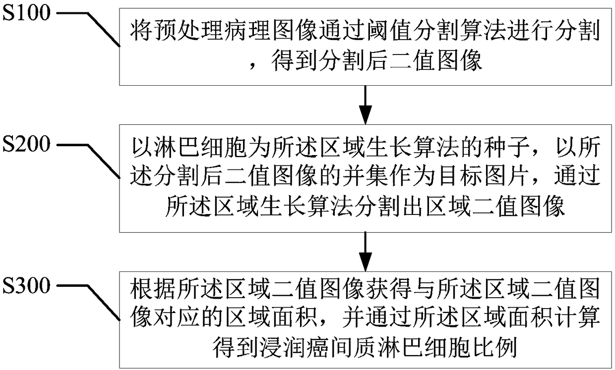
[0062] refer to figure 2 , the first embodiment of the present invention provides a breast cancer image recognition method, including:

[0063] Step S100, segmenting the preprocessed pathological image through a threshold segmentation algorithm to obtain a segmented binary image;

[0064] As mentioned above, it needs to be understood that a binary image (Binary Image) means that each pixel on the image has only two possible values ​​​​or grayscale states. People often use black and white, B&W, and monochrome images to represent binary images. . A binary image means that in the image, there are only two gray levels, that is, any pixel in the image is either 0 or 1, and there is no other transitional gray value.

[0065] As mentioned above, it needs to be understood that image segmentation is the technology and process of dividing an image into several specific regions with unique properties and proposing objects of interest. It is a key step from image processing to image a...

Embodiment 2

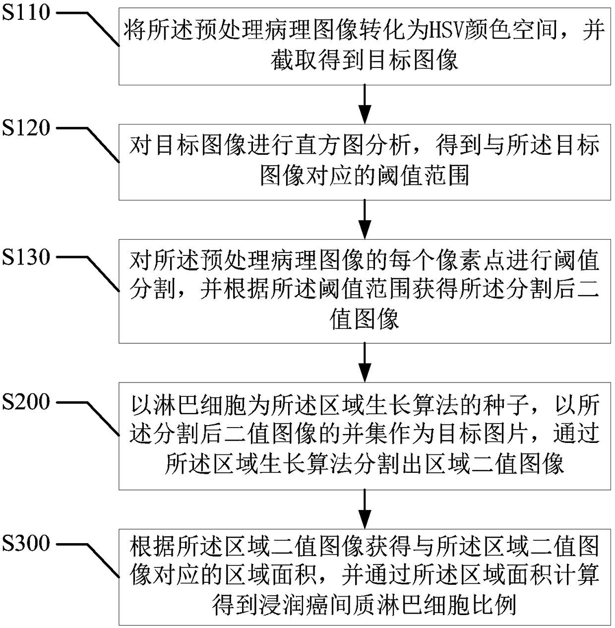
[0076] refer to image 3 , the second embodiment of the present invention provides a breast cancer image recognition method, based on the above figure 2 In the first embodiment shown, the step S100 of "segmenting the preprocessed pathological image through a threshold segmentation algorithm to obtain a segmented binary image" includes:

[0077] Step S110, converting the preprocessed pathological image into HSV color space, and intercepting and obtaining the target image;

[0078] As mentioned above, it needs to be understood that HSV (Hue, Saturation, Value) is a color space created by A.R. Smith in 1978 based on the intuitive characteristics of color, also known as the Hexcone Model (Hexcone Model). The parameters of the color in this model are: hue (H), saturation (S), and lightness (V).

[0079] As mentioned above, the hue H is measured by angle, and the value ranges from 0° to 360°. It is calculated counterclockwise from red, red is 0°, green is 120°, and blue is 240°. ...

Embodiment 3

[0092] refer to Figure 4 , the third embodiment of the present invention provides a breast cancer image recognition method, based on the above image 3 In the second embodiment shown, the step S130 of "performing threshold segmentation on each pixel of the preprocessed pathological image, and obtaining the segmented binary image according to the threshold range" includes:

[0093] Step S131, performing threshold segmentation on each pixel of the preprocessed pathological image, and confirming the pixels whose pixel values ​​in the three dimensions of H, S, and V are all within the threshold range;

[0094] Step S132, set the pixel points whose pixel values ​​in the three dimensions of H, S, and V are all within the threshold range to 1, and set the pixel values ​​in the three dimensions of H, S, and V that are not all in the threshold range Set the pixel points inside to 0 to obtain the segmented binary image.

[0095] As mentioned above, perform threshold segmentation on e...


PUM


Abstract

The invention provides a breast cancer image identification method and device, and a user terminal. The method comprises the following steps of using a threshold segmentation algorithm to segment a preprocessing pathology image and acquiring segmented binary images; taking lymphocytes as the seed of a region growing algorithm, taking the union set of the segmented binary images as a target picture, and using the region growing algorithm to segment and acquiring area binary images; and acquiring an area corresponding to the area binary images according to the area binary images, and calculatingand acquiring the proportion of infiltrating carcinoma interstitial lymphocytes through the area. By using the method of the invention, the identification of cancer cells in the pathology image of abreast cancer through a computer is realized, the accuracy of breast cancer pathology image diagnosis is increased, the errors of artificial naked eye determination are avoided, breast cancer pathology image diagnosis time is shortened, diagnosis efficiency is increased and convenience is brought for the diagnosis work of a doctor.

Description

technical field [0001] The present invention relates to the technical field of image recognition, and more specifically, relates to a breast cancer image recognition method, device and user terminal. Background technique [0002] In medicine, cancer refers to malignant tumors originating from epithelial tissue, and is the most common type of malignant tumors. A cancer cell is a mutated cell. It is the source of cancer. Cancer cells are different from normal cells. They have three characteristics: infinite proliferation, transformation and easy transfer. They can infinitely proliferate and destroy normal cell tissues. In addition to uncontrollable division of cancer cells (capable of multipolar division), cancer cells can also partially invade surrounding normal tissues and even transfer to other parts of the body through the internal circulatory system or lymphatic system. [0003] Generally, in most cancer diagnoses (including breast cancer), doctors need to make a diagno...

Claims


Application Information

Patent Timeline
Patent Type & Authority Applications(China)
IPC IPC(8): G06T7/00G06T7/11G06T7/155G06T7/187G06T7/62G06T5/30
CPCG06T5/30G06T7/0012G06T7/11G06T7/155G06T7/187G06T7/62G06T2207/30068G06T2207/30096
Inventor 衣杨吴昱焜张念旭谢韬李仲泓周翼丰
Owner SUN YAT SEN UNIV
Why Eureka
Try Eureka