Ultrasonic image processing method, device, storage medium, and ultrasonic imaging device

An ultrasonic image and ultrasonic imaging technology, applied in the field of medical image processing, can solve the problems of unclear depth range and cumbersome operation, and achieve the effect of simplifying operation and improving adjustment accuracy

Active Publication Date: 2018-10-16
SONOSCAPE MEDICAL CORP
View PDF6 Cites 6 Cited by
  • Summary
  • Abstract
  • Description
  • Claims
  • Application Information

AI Technical Summary

Problems solved by technology

[0004] However, the inventor found that when the TGC parameter adjustment needs to be performed after the ultrasonic image is enlarged, since the currently displayed ultrasonic image is a part of the ultrasonic image before the enlargement, the user needs to select from a plurality of TGC parameter adjustment keys that are compatible with the currently displayed The TGC parameter adjustment key corresponding to the depth to which the ultrasonic image belongs is used for adjustment, and since the user does not know which depth range of the ultrasonic image before zooming in corresponds to the currently displayed ultrasonic image, it takes the user several attempts to find the corresponding TGC parameter adjustment key, making the user's operation more cumbersome

Method used


Image

  • Ultrasonic image processing method, device, storage medium, and ultrasonic imaging device
  • Ultrasonic image processing method, device, storage medium, and ultrasonic imaging device
  • Ultrasonic image processing method, device, storage medium, and ultrasonic imaging device

Examples


Embodiment Construction

[0041] The following will clearly and completely describe the technical solutions in the embodiments of the present invention with reference to the accompanying drawings in the embodiments of the present invention. Obviously, the described embodiments are only some, not all, embodiments of the present invention. Based on the embodiments of the present invention, all other embodiments obtained by persons of ordinary skill in the art without making creative efforts belong to the protection scope of the present invention.

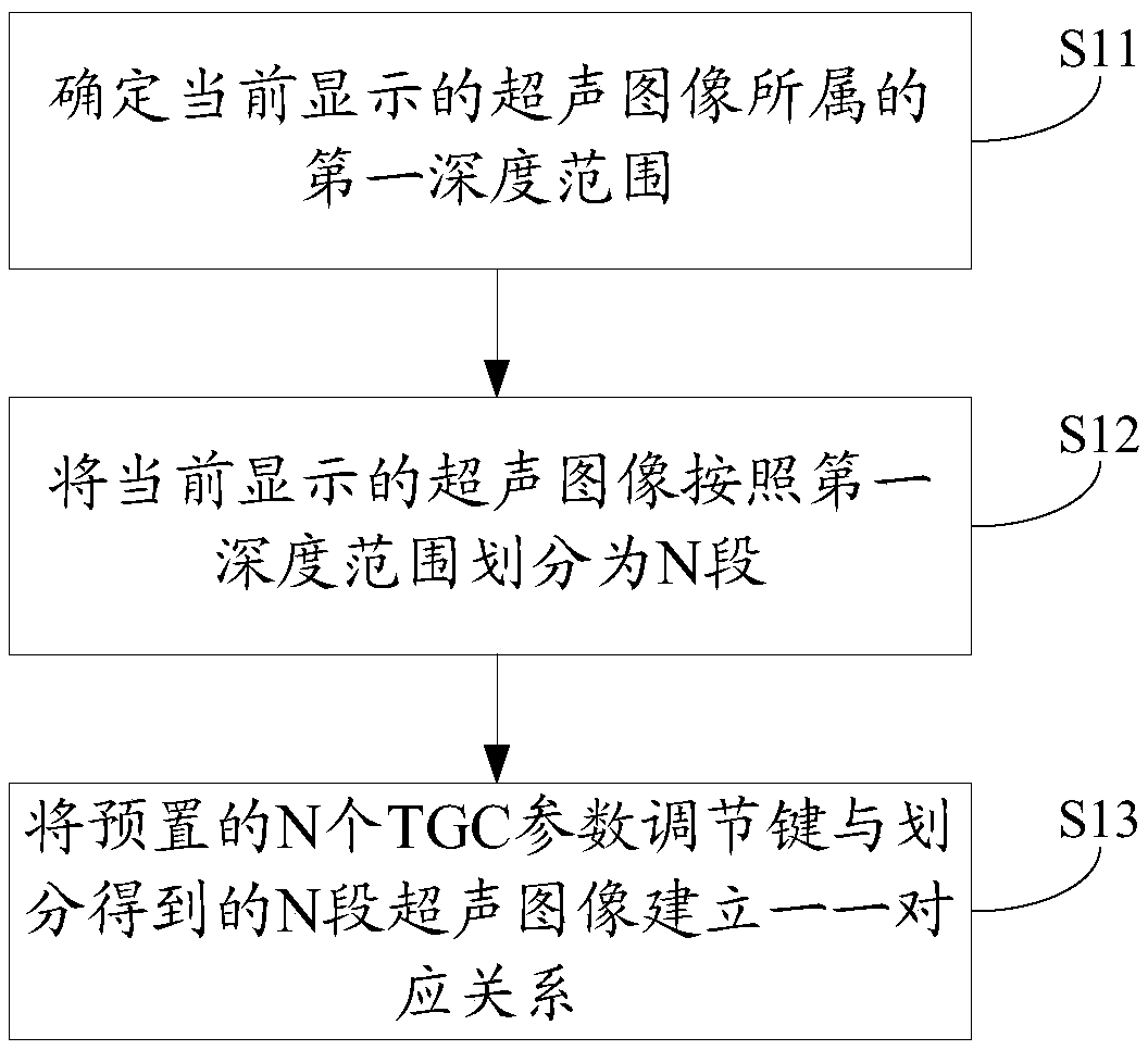
[0042] see figure 1 , figure 1 An implementation flowchart of the ultrasonic image processing method provided in the embodiment of the present application may include:

[0043] Step S11: Determine the depth range to which the currently displayed ultrasound image belongs (for convenience of description, it is recorded as the first depth range).

[0044] The first depth range is at least a part of the maximum depth range scanned by the ultrasound imaging devic...


PUM


Abstract

The embodiment of the invention discloses an ultrasonic image processing method, a device, a computer-readable storage medium and an ultrasonic imaging device. Regardless of the depth range of the currently displayed ultrasound image, the currently displayed ultrasound image is divided into N segments according to the depth range; a preset N-time gain compensation parameter adjustment key is usedfor establishing a one-to-one relation with the divided N-segment ultrasound images, when the currently displayed ultrasound image is a local image, any one of the above-mentioned N time gain compensation parameter adjustment keys can adjust the currently displayed ultrasound image so that the user can approximate the time gain compensation parameter adjustment key corresponding to the image of the depth to be adjusted by visual inspection instead of a blind attempt, accordingly the operation of the user is simplified, and the adjusting precision is improved.

Description

technical field [0001] The present application relates to the technical field of medical image processing, and more specifically, to an ultrasonic image processing method, device, computer-readable storage medium, and ultrasonic imaging equipment. Background technique [0002] Ultrasound images are images commonly used in the medical field. When the user (usually a medical worker) uses the ultrasonic imaging device to scan the irradiated object, the image displayed by the ultrasonic imaging device (hereinafter referred to as the ultrasonic image) is an image of a section of the irradiated object at different depths. . [0003] In practical applications, users sometimes need to zoom in on ultrasound images to view details. In the enlarged image, it may also be necessary to adjust a time gain compensation (Time Gain Compensate, TGC) parameter, so that the display effect of the ultrasound image can better meet the needs of the user. At present, for the convenience of viewing...

Claims


Application Information

Patent Timeline
Patent Type & Authority Applications(China)
IPC IPC(8): A61B8/00
CPCA61B8/4411A61B8/461A61B8/469A61B8/5215
Inventor 许龙何丹妮廖静秋
Owner SONOSCAPE MEDICAL CORP
Why Eureka
Try Eureka