Image segmentation method, image segmentation apparatus and electronic device

A technology of image segmentation and sample images, applied in the field of image processing, can solve problems such as inaccurate segmentation of brain parenchyma

Active Publication Date: 2018-11-06
SHENZHEN BRAINNOW MEDICAL TECH CO LTD
View PDF4 Cites 8 Cited by
  • Summary
  • Abstract
  • Description
  • Claims
  • Application Information

AI Technical Summary

Problems solved by technology

[0005] In view of this, the present application provides an image segmentation method, an image segmentation device, electronic equipment, and a computer-readable storage medium, which can solve the technical problem of inaccurate brain parenchyma segmentation in the prior art

Method used


Image

  • Image segmentation method, image segmentation apparatus and electronic device
  • Image segmentation method, image segmentation apparatus and electronic device
  • Image segmentation method, image segmentation apparatus and electronic device

Examples


Embodiment 1

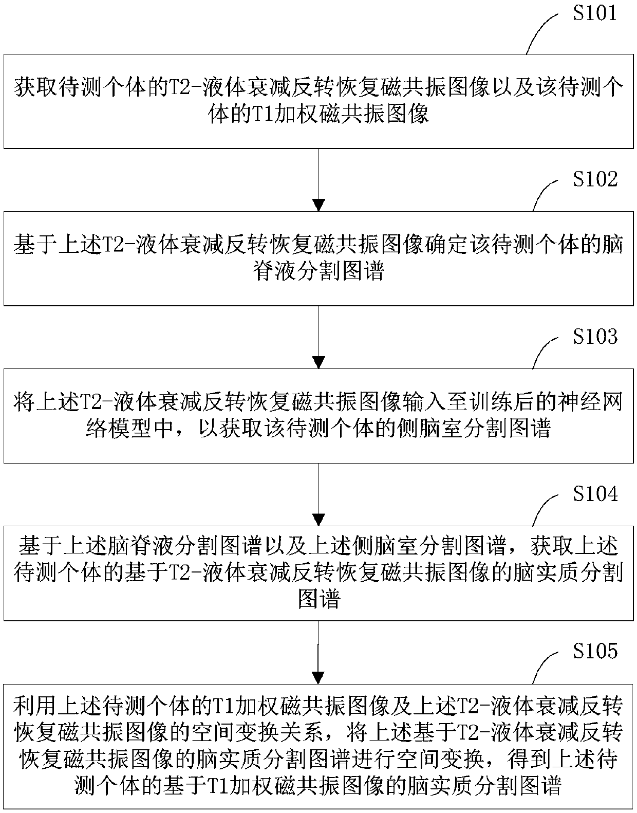
[0034] The image segmentation method provided by Embodiment 1 of the present application is described below, please refer to the attached figure 1 , the image segmentation method in the embodiment of the present application includes steps S101-S105:

[0035] In step S101, acquire the T2-fluid attenuated inversion recovery magnetic resonance image of the individual to be tested and the T1 weighted magnetic resonance image of the individual to be tested;

[0036] In the embodiment of the present application, it is necessary to obtain the T2-fluid attenuated inversion recovery (T2-fluidattenuated inversion recovery, T2-FLAIR) magnetic resonance image of the individual to be tested first and the T1-weighted magnetic resonance image of the individual to be tested, so as to facilitate subsequent Based on the above-mentioned T2-FLAIR magnetic resonance image and T1 weighted magnetic resonance image, the brain parenchyma segmentation map based on the T1 weighted magnetic resonance ima...

Embodiment 2

[0060] Another image segmentation method provided by Embodiment 2 of the present application is described below, please refer to the attached Image 6 , the image segmentation method of Embodiment 2 of the present application includes steps S201-S205:

[0061] In step S201, acquire the T2-fluid attenuated inversion recovery magnetic resonance image of the individual to be tested and the T1 weighted magnetic resonance image of the individual to be tested;

[0062] In step S202, the cerebrospinal fluid segmentation atlas of the individual to be tested is determined based on the above-mentioned T2-fluid attenuated inversion recovery magnetic resonance image;

[0063] In this embodiment of the present application, the above steps S201 and S202 are performed in the same manner as steps S101 and S102 in Embodiment 1. For details, please refer to the description of Embodiment 1, which will not be repeated here.

[0064] In step S203, based on the spatial transformation relationship ...

Embodiment 3

[0076] Embodiment 3 of the present application provides an image segmentation device. For the convenience of description, only the parts related to the present application are shown, such as Figure 7 As shown, the image segmentation device 300 includes:

[0077] An image acquisition module 301, configured to acquire the T2-fluid attenuated inversion recovery magnetic resonance image of the individual to be tested and the T1 weighted magnetic resonance image of the individual to be tested;

[0078] A cerebrospinal fluid segmentation module 302, configured to determine the cerebrospinal fluid segmentation atlas of the individual to be tested based on the T2-fluid attenuated inversion recovery magnetic resonance image;

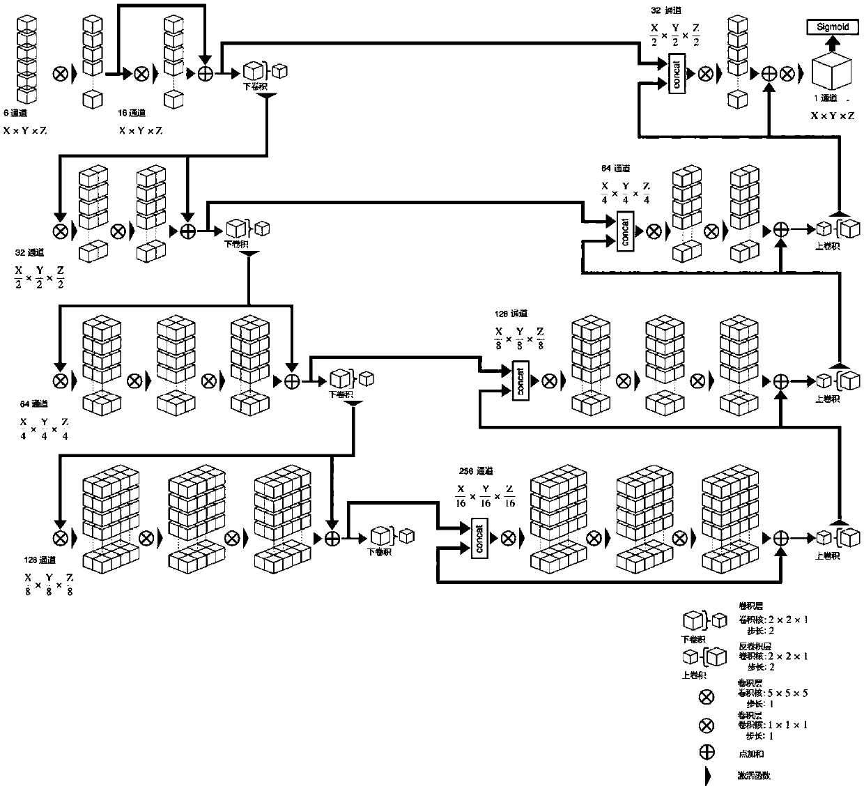
[0079] The lateral ventricle acquisition module 303 is configured to input the T2-fluid attenuated inversion recovery magnetic resonance image into the trained neural network model to obtain the lateral ventricle segmentation map of the individual to be tested, ...


PUM


Abstract

The invention provides an image segmentation method, an image segmentation apparatus and an electronic device. The method comprises the steps of acquiring a T2-fluid attenuated inversion recovery magnetic resonance image and a T1 weighted magnetic resonance image of a to-be-tested individual; based on the T2-fluid attenuated inversion recovery magnetic resonance image, determining a cerebrospinalfluid segmentation graph; inputting the T2-fluid attenuated inversion recovery magnetic resonance image to a neural network model, thereby obtaining a lateral ventricle segmentation graph; on the basis of the cerebrospinal fluid segmentation graph and the lateral ventricle segmentation graph, obtaining a brain parenchyma segmentation graph based o the T2-fluid attenuated inversion recovery magnetic resonance image; and by utilizing a spatial conversion relationship between the T1 weighted magnetic resonance image and the T2-fluid attenuated inversion recovery magnetic resonance image, obtaining a brain parenchyma segmentation graph based on the T1 weighted magnetic resonance image. According to the technical scheme, a cerebrospinal fluid part and a non-cerebrospinal fluid part can be moreaccurately divided, so that the more accurate brain parenchyma segmentation graphs can be obtained.

Description

technical field [0001] The present application belongs to the technical field of image processing, and in particular relates to an image segmentation method, an image segmentation device, electronic equipment, and a computer-readable storage medium. Background technique [0002] Currently, the prevalence of multiple sclerosis is increasing and it is affecting the global population between the ages of 20 and 80. Multiple sclerosis is caused by the disorder of the human immune system. Immune cells mistakenly attack the myelin basic protein that makes up the insulating sheath of nerve tissue. Multiple sclerosis will eventually lead to the loss of motor function of the patient or cause symptoms such as brain dementia. Great pain for patients and families. [0003] Under normal circumstances, multiple sclerosis will affect the operation of white matter, gray matter and ventricular system to change the volume of the brain parenchyma. Although the volume of the brain parenchyma of...

Claims


Application Information

Patent Timeline
Patent Type & Authority Applications(China)
IPC IPC(8): G06T7/168G06N3/08
CPCG06N3/08G06T2207/10088G06T2207/30016G06T7/168
Inventor 罗怡珊
Owner SHENZHEN BRAINNOW MEDICAL TECH CO LTD
Why Eureka
Try Eureka