Method and system for medical image automatic segmentation, apparatus and storage medium
A medical image and automatic segmentation technology, applied in the application field of computer analysis technology, can solve the problems of increasing segmentation difficulty, lack of universality and robustness, and influence of image data, reducing information processing capacity, improving classification performance, The effect of accurate segmentation
- Summary
- Abstract
- Description
- Claims
- Application Information
AI Technical Summary
Problems solved by technology
Method used
Image

Examples
Embodiment 1
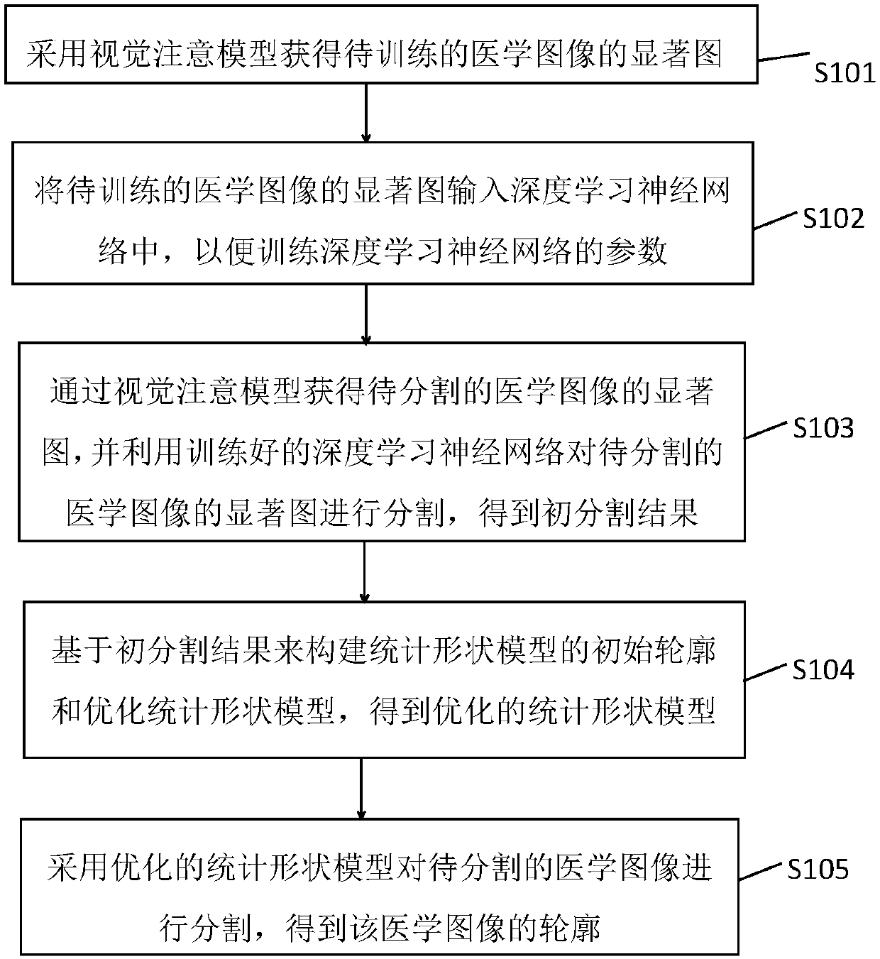
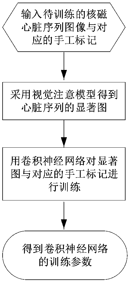
[0049] figure 1 It is a schematic flow chart of the automatic segmentation method for medical images provided in Embodiment 1 of the present invention. The execution subject of the automatic segmentation method provided in the embodiment of the present invention may be the automatic segmentation system provided in the embodiment of the present invention, and the system can be integrated into a mobile terminal Devices (for example, smart phones, tablet computers, notebooks, etc.) can also be integrated into the server, and the automatic segmentation system can be implemented by hardware or software. The automatic segmentation method provided by the embodiment of the present invention is particularly suitable for the situation of computer-aided diagnosis of cardiac images based on nuclear magnetic images, which will be described below in conjunction with the embodiments.
[0050] Such as figure 1 As shown, the automatic segmentation method specifically includes:
[0051] S101,...
Embodiment 2
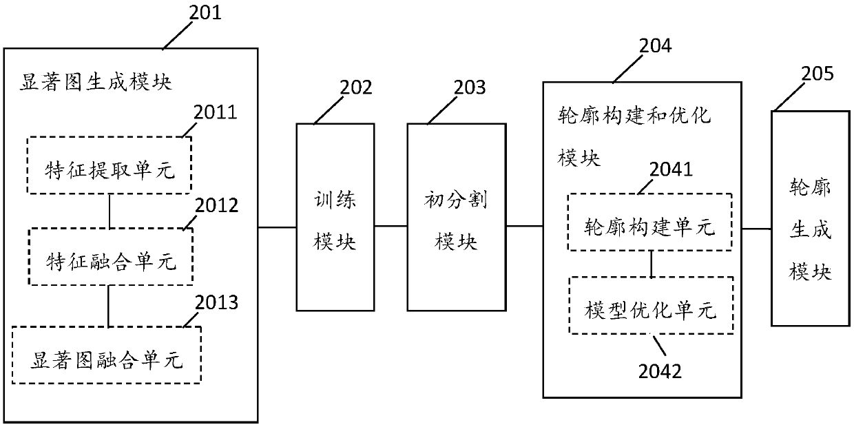
[0062] figure 2 It is a schematic structural diagram of the automatic segmentation system of medical images provided by Embodiment 2 of the present invention. The system can be integrated into mobile terminal equipment (such as smart phones, tablet computers, notebooks, etc.), and can also be integrated into servers. The positioning device can Implemented in hardware or software.
[0063] Such as figure 2 As shown, the system specifically includes a saliency map generation module 201, a training module 202, an initial segmentation module 203, a contour construction and optimization module 204, and a contour generation module 205;
[0064] The saliency map generation module 201 adopts the visual attention model to obtain the saliency map of the medical image to be trained;
[0065] The training module 202 is used to input the saliency map of the medical image to be trained into the deep learning neural network, so as to train the parameters of the deep learning neural netwo...
Embodiment 3
[0151] Figure 11 It is a schematic structural diagram of the device provided in Embodiment 3 of the present invention, and the device can be used to realize the automatic segmentation method of medical images according to the embodiment of the present invention.
[0152] exist Figure 11 Among them, a central processing unit (CPU) 601 executes various processes according to programs stored in a read only memory (ROM) 602 or programs loaded from a storage section 608 to a random access memory (RAM) 603 . In the RAM 603, data required when the CPU 601 executes various processes and the like is also stored as necessary. The CPU 601 , ROM 602 , and RAM 603 are connected to each other via a bus 604 . The input / output interface 605 is also connected to the bus 604 .
[0153] The following components are also connected to the input / output interface 605: an input section 606 (including a keyboard, a mouse, etc.), an output section 607 (including a display such as a cathode ray tub...
PUM
 Abstract
Description
Claims
Application Information